Neutrophil extracellular traps regulate the development of innate and adaptive immune system




















DOI: 10.32364/2587-6821-2020-4-1-38-41
A.N. Kazimirskii, J.M. Salmasi, G.V. Poryadin
Pirogov Russian National Research Medical University, Moscow, Russian Federation
Neutrophils are the main constitutes responsible for innate immunity. They are important for early anti-infectious protection. However, the interaction between innate and adaptive immune system in early inflammation still remains elusive. There is no general conception of human innate and adaptive immune system interaction. We suggest to examine the interaction between innate and adaptive immune system when pathogens enter the body as several consecutive steps. The first step is pathogen-induced neutrophil activation. At this stage, neutrophils produce and release interferon gamma and activate the synthesis of NADPH oxidase, myeloperoxidase, and arginase. This results in the activation of innate immune response by attracting cytotoxic T cells and natural killer cells to the site of inflammation. The second step is the generation of neutrophil extracellular traps (NETs) which bind, inactivate, or damage pathogens by reactive oxygen nitrogen species. This stage involves the inhibition of adaptive immune response due to the local decrease in arginine concentration leading to reduced T cell functionality. The third step of acute infectious inflammation is downregulation of innate immune response receptors (TLRs) mediated by toll interacting protein. As a result, the reactivity of innate immune system decreases. NET release is blocked. The arrest of arginine release from neutrophils promotes antigen presentation and adaptive immune response activation. Inconsistency or deficiency of neutrophil activation steps result in the complications of inflammation.
Keywords: neutrophil extracellular traps, gamma-interferon, mitochondrial antiviral-signaling protein (MAVS), TOLLIP, innate immunity, adaptive immunity.
For citation: Kazimirskii A.N., Salmasi J.M., Poryadin G.V. Neutrophil extracellular traps regulate the development of innate and adaptive immune system. Russian Medical Review. 2020;4(1):38–41. DOI: 10.32364/2587-6821-2020-4-1-38-41.
Background
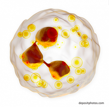
Neutrophils are the largest pool of leukocytes that contribute to local immune reactions and the main constitutes responsible for innate immunity. These cells represent the first line of the defense against inflammation and infections. Receptors of innate immune cells interact with potentially dangerous exogenous and endogenous ligands. Invading pathogens induce inflammatory reaction that attracts neutrophils from blood to tissues. At the site of inflammation, neutrophils facilitate killing microorganisms in several ways, largely by phagocytosis, antimicrobial substance release, and neutrophil extracellular trap (NET) formation. NET formation, or NETosis, is the mechanism used by neutrophils to destroy microorganisms [1, 2]. The interaction between neutrophils and microorganisms results in cell activation and the release of NETs composed of decondensed DNA, cytosolic proteins, enzymes, granular proteins, and histones. This complex provides a rather high local concentration of antimicrobial substances to inactivate and kill pathogens irrespective of their ensnaring by neutrophils. Even though the mechanism of NET formation is still understudied, it is known that neutrophilic NADPH oxidase activity contributes to this process. Only few studies describe NETs that function without preliminary activation of NADPH oxidase. However, antibacterial efficacy of these NETs is predictably low. It was demonstrated that contact interactions between activated neutrophils and apoptotic cells are an important factor that initiates NET deployment. Moreover, neutrophils containing residual apoptotic cells do not release NETs but activate phagocytosis [3].
In addition to antimicrobial properties, NETs create a barrier preventing pathogens from spreading. A myriad of studies on the role of NETs in infectious inflammation have demonstrated that both deficient and excessive formation of NETs is unfavorable for inflammation. Deficient NET formation is accounted for by congenital factors but may be induced by pathogens as well. Excessive NET formation results in tissue damage by reactive oxygen species (ROS) and peptides generating pores (e.g., LL-37), blood coagulation, thromboses, and hemophagy. Studies emerged in recent years demonstrate important protective role of NETs that involves both direct killing of microbes and regulating the interactions between innate and adaptive immunity [4].
Role of NETs in host antiviral defense
Viral infection induces the generation of mitochondrial antiviral-signaling protein (MAVS) aggregates. MAVS characterizing by prion-like activity [5] actively stimulates immune signaling. Autophagy is important for MAVS-mediated signaling. Following viral invasion ubiquitin ligase (RNF34) binds with MAVS of mitochondrial membranes and promotes autophagic MAVS degradation required for the regeneration of damaged mitochondria in viral infections. RNF34-mediated autophagic MAVS degradation regulates innate immunity and mitochondrial homeostasis and controls cellular infection [6]. Intracellular reactions are induced after the recognition of viral products via pathogen-associated molecular patters. This recognition triggers signaling cascades that activate intracellular innate immune defense and inflammatory response that, in turn, facilitates adaptive immunity. Activated signals from RIG-1 cooperate with adaptor protein MAVS to initiate signaling cascade that stimulates transcription factors IRF3 and NF-κB. This results in the expression of antiviral gene products and production of type I and III interferons (IFNs) that reduce the number of viral copies in an infected cell and surrounding tissues [7].
Innate immune response to a pathogen has several steps. First, infected macrophages and dendritic cells recruit and activate neutrophils by producing and secreting IFN type I. At this stage, pathogen interacts with innate immune response receptors (TLRs) or intracellular receptors RLR (RIG-I, MDA5, LGP2). A group of RLR receptors function as sensors of viral replication in the cytoplasm and detect viral replication via direct interaction with double-stranded RNA of viral genome. Peripheral blood neutrophils reach the site of inflammation, become activated by IFN-I (IFN-α and IFN-β), and produce IFN-γ. IFN-γ attracts and activates NK cells and cytotoxic CD8+ T cells that are able to recognize (when having specific receptors) and kill infected cells. The development of innate immune reactions is accompanied by IFN-γproduction by peripheral blood neutrophils and enhanced functional activity of cytotoxic cells.
Experimental findings demonstrate that MAVS can increase its expression and the production of antiviral and anti-inflammatory cytokines and induce apoptosis [8]. Contact interactions between activated neutrophils and infected cells undergoing apoptosis contribute to NET formation followed by phagocytosis and hydrolysis of all intracellular components of infected cells (including viral DNA or RNA) [3, 9].
These data suggest nonspecific antiviral host defense associated with NET formation. Membrane protein MAVS located in the outer mitochondrial membrane is the main sensor of this system. Binding with viral DNA or RNA results in specific conformational changes that, through a number of steps, induce apoptosis of infected cells. Contact interactions between neutrophils and infected cells undergoing apoptosis result in the formation of NETs that ensnare apoptotic cells and provide phagocytosis and hydrolysis of all cellular and viral structures.
Role of NETs in the regulation of immune response
NET deployment after the interaction with pathogen occurs when achieving a certain level of neutrophil activation involving genome expression and production of anti-infective defense enzymes, cytokines, and regulatory proteins including Toll-interacting protein (Tollip). Tollip is an ubiquitin-binding protein that regulates (limits) innate immune response including Toll-like receptor (TLR) signaling pathways and is the key negative regulator of innate immunity preventing excessive inflammation. Its role was studied in patients with various diseases and experimental animals. Thus, reduced blood levels of Tollip were revealed in patients with severe inflammatory disorders (i.e., septic colitis with unfavorable outcome) as compared with healthy persons. Tollip-deficient neutrophils were characterized by the reduced migration in response to the powerful chemoattractant N-formyl-met-leu-phe (bacterial protein degradation product), reduced potentialities of NET formation, and reduced activity to kill bacteria [10].
Tollip deficiency is associated with the increased risk of tuberculosis. Tollip polymorphisms were found in these patients thus demonstrating the role of the mechanism of negative regulation of TLR signaling in the pathogenesis of human tuberculosis [11]. Tollip deficiency induces rapid involvement of adaptive immune system in the response. Tollip-deficient neutrophils significantly enhance T cell activation via upregulation of co-stimulating molecule CD80 and downregulation of inhibiting molecule PD-L1. The lack of Tollip increases STAT5 generation and decreases STAT1 generation; these transcription factors are responsible for the expression of CD80 and PD-L1, respectively [12]. These data illustrate the important role of Tollip in innate immune reactions. As NETs are deployed, Tollip inhibits TLR expression thus preventing the interaction between neutrophils and pathogens. Meanwhile, NET formation becomes slower thus representing the mechanism that limits innate immunity at early stages of inflammation. Impairment of this process results in the continuous release of enzymes, ROS, and biologically active substances from NETs and promote tissue damage.
Therefore, physiological role of Tollip is to maintain a balance between innate and adaptive immune reactions. Enzymes generated during NET activation are among other important factors affecting immunity.
NADPH oxidase, myeloperoxidase, and arginase are major secreted enzymes reaching the site of inflammation after NET deployment. The physiological role of NADPH oxidase and myeloperoxidase is to generate oxygen and nitrogen radicals characterized by robust anti-infective effects while the role of arginase is to inhibit adaptive immunity [13]. Numerous studies demonstrate that these pre-activated enzymes enter extracellular space in early inflammation while the impairment of this process is always detrimental. This step is essential for the interaction between pathogens and host immune system but limited in time. Physiological limiter of NET formation is Tollip. The importance of arginase that reaches the site of inflammation being released from activated neutrophils in the course of NET formation and resulting inhibition of adaptive immunity remains elusive and is interesting for the research community.
The association between blood arginase activity, neutrophil/lymphocyte ratio, and disease severity was found in acute ischemic stroke. In addition, increased amount of arginase-1 mRNA and soluble surface CD100 receptor localized on endothelial cells was discovered 24 hours after brain injury. These findings demonstrate neutrophil activation and the involvement of vascular endothelium in inflammation. Moreover, increased levels of matrix metalloproteinase-9 (MMP-9) and myeloid differentiation factor-2 (MD-2) were detected in these patients. MMP-9 contributes to extracellular matrix remodeling while MD-2 is a glycoprotein that binds extracellular domain of innate immune receptor (TLR4) and activates it thus playing an important role in the innate immune response [14, 15]. Acute stroke modifies systemic immune response in peripheral blood, however, molecular mechanism upregulating arginase expression is an enigma. Examination of patients with acute stroke has demonstrated that microRNA miR-340-5p binds with 3'-untranslated region of arginase-1 gene and suppresses arginase gene expression. Decrease in miR-340-5p levels found in acute stroke is suggested to abolish the inhibition of arginase-1 gene expression that eventually results in its extensive synthesis [16]. The ability of murine neutrophils to release arginase from preformed granules was demonstrated in the murine model of temporary occlusion of the middle cerebral artery. Reduced expression of CD3 ζ-chain (CD3ζ) on T cells was found being in line with the decrease in their functionality [17]. Acute infectious processes result in rapid upregulation of arginase activity in peripheral blood. Significant increase in the activity of peripheral blood arginase was revealed in children with necrotic enterocolitis. Moreover, it was demonstrated that this enzyme is produced by infiltrating neutrophils [18]. During acute human infections, the number of peripheral blood neutrophils synthesizing arginase is proportional to disease severity. These neutrophils affects CD3ζ expression and T cell function thus contributing to T cell dysfunction observed in sepsis [19, 20]. Chronicity of inflammation is also associated with increased arginase activity at the site of inflammation. Tuberculous granulomas (representing compact organized aggregations of infected and non-infected macrophages, T cells, neutrophils, and other cells) are characterized by the increased activity of arginase released by macrophages. Induction of arginase arrests T cell proliferation thus depriving them of their functionality [21].
These findings illustrate that the interaction between innate and adaptive immunity demonstrate a certain reciprocity at the early stage of acute inflammation. These findings also show the inhibition of adaptive immunity at the early stage of acute inflammation associated with NET formation. During NET formation, arginase enters extracellular space while arginine concentration reduces. The result is the suppression of T cell functional activity and the inhibition of antigen presentation. This step is characterized by robust activation of innate immunity and simultaneous deactivation of adaptive immunity. As innate immune receptor expression downregulates, innate immune system reactivity decreases under the effect of Tollip. Antigen presentation in the lack of arginine release from neutrophils promote activation of adaptive immunity.
Conclusions
The analysis of experimental findings has highlighted previously unknown NET effects and has demonstrated their importance for the development of antiviral immunity and the involvement in the regulation of innate and adaptive immunity.
Pathogen-induced neutrophil activation occurs at the early stage of acute infectious inflammation. At this stage, neutrophils produce and release IFN-γ and stumulate the synthesis of NADPH oxidase, myeloperoxidase, and arginase. A certain high level of NADPH oxidase activity initiate the formation of NETs that bind, inactivate, or damage pathogens using reactive oxygen and nitrogen species. During NET deployment, arginase enters extracellular space while arginine concentration reduces thus resulting in the suppression of T cell functionality. Further development of acute infectious inflammation is accompanied by the downregulation of TLR expression mediated by Tollip. As a result, NET deployment is curtailed. The upregulation of T cell functionality in the lack of arginine release from neutrophils promote the activation of adaptive immune response.
About the authors:
Aleksandr N. Kazimirskii — PhD (Biology), Associate Professor, leading researcher of the Division of Molecular Technologies, ORCID iD 0000-0002-3079-4089;
Jean M. Salmasi — MD, PhD, Professor, Head of the Department of Pathophysiology and Clinical Pathophysiology, ORCID iD 0000-001-8524-0019;
Gennadiy V. Poryadin — MD, PhD, Professor, Corresponding Member of RAS, Department of Pathophysiology and Clinical Pathophysiology, ORCID iD 0000-0003-2010-3296.
Pirogov Russian National Research Medical University. 1, Ostrovityanov str., Moscow, 117437, Russian Federation.
Contact information: Aleksandr N. Kazimirskii, e-mail: alnica10@mail.ru.
Financial Disclosure: no authors have a financial or property interest in any material or method mentioned. There is no conflict of interests. Received 24.01.2020.
2. Fuchs T.A., Abed U., Goosmann C. et al. Novel cell death program leads to neutrophil extracellular traps. J Cell Biol. 2007;176(2):231–241. DOI: 10.1083/jcb.200606027.
3. Manfredi A.A., Ramirez G.A., Rovere-Querini P., Maugeri N. The Neutrophil’s Choice: Phagocytose vs Make Neutrophil Extracellular Traps. Front Immunol. 2018;9:288. DOI: 10.3389/fimmu.2018.00288. eCollection 2018.
4. Kazimirskii A.N., Salmasi J.M., Poryadin G.V. Coordination of Innate and Adaptive Immunity Depending on Neutrophilic Extracellular Traps Formation. Austin J Clin Immunol. 2019;6(1):1037.
5. Cai X., Xu H., Chen Z.J. Prion-Like Polymerization in Immunity and Inflammation. Cold Spring Harb Perspect Biol. 2017;9(4): pii: a023580. DOI: 10.1101/cshperspect.a023580.
6. He X., Zhu Y., Zhang Y. et al. RNF34 functions in immunity and selective mitophagy by targeting MAVS for autophagic degradation. EMBO J. 2019;38(14): e100978. DOI: 10.15252/embj.2018100978.
7. Kell A.M., Gale M. Jr. RIG-I in RNA virus recognition. Virology. 2015;479–480:110–121. DOI: 10.1016/j.virol.2015.02.017.
8. Dong W., Lv H., Li C. et al. MAVS induces a host cell defense to inhibit CSFV infection. Arch Virol. 2018;163(7):1805–1821. DOI: 10.1007/s00705-018-3804-z.
9. Hamam H.J., Palaniyar N. Histone Deacetylase Inhibitors Dose-Dependently Switch Neutrophil Death from NETosis to Apoptosis. Biomolecules. 2019;9(5): pii: E184. DOI: 10.3390/biom9050184.
10. Diao N., Zhang Y., Chen K. et al. Deficiency in Toll-interacting protein (Tollip) skews inflamed yet incompetent innate leukocytes in vivo during DSS-induced septic colitis. Sci Rep. 2016;6:34672. DOI: 10.1038/srep34672.
11. Shah J.A., Vary J.C., Chau T.T.H. et al. Human TOLLIP Regulates TLR2 and TLR4 Signaling and Its Polymorphisms Are Associated with Susceptibility to Tuberculosis. J Immunol. 2012;189(4):1737–1746. DOI: 10.4049/jimmunol.1103541.
12. Zhang Y., Lee C., Geng S., Li L. Enhanced tumor immune surveillance through neutrophil reprogramming due to Tollip deficiency. JCI Insight. 2019;4(2): pii:122939. DOI: 10.1172/jci.insight.122939.
13. Kazimirskii A.N., Poryadin G.V., Salmasi Z.M., Semenova L.Y. Endogenous Regulators of the Immune System (sCD100, Malonic Dialdehyde, and Arginase). Bull Exp Biol Med. 2018;164(5):693–700. DOI: 10.1007/s10517-018-4061-6.
14. Petrone A.B., O’Connell G.C., Regier M.D. et al. The Role of Arginase 1 in Post-Stroke Immunosuppression and Ischemic Stroke Severity. Transl Stroke Res. 2016;7(2):103–110. DOI: 10.1007/s12975-015-0431-9.
15. Petrone A.B., Gionis V., Giersch R., Barr T.L. Immune biomarkers for the diagnosis of mild traumatic brain injury. NeuroRehabilitation. 2017;40(4):501–508. DOI: 10.3233/NRE-171437.
16. Yoo H., Kim J., Lee A.R. et al. Alteration of microRNA 340–5p and Arginase-1 Expression in Peripheral Blood Cells during Acute Ischemic Stroke. Mol Neurobiol. 2019;56(5):3211–3221. DOI: 10.1007/s12035-018-1295-2.
17. Sippel T.R., Shimizu T., Strnad F. et al. Arginase I release from activated neutrophils induces peripheral immunosuppression in a murine model of stroke. J Cereb Blood Flow Metab. 2015;35(10):1657–1663. DOI: 10.1038/jcbfm.2015.103.
18. Leung K.T., Chan K.Y., Ma T.P. et al. Dysregulated expression of arginine metabolic enzymes in human intestinal tissues of necrotizing enterocolitis and response of CaCO2 cells to bacterial components. J Nutr Biochem. 2016;29:64–72. DOI: 10.1016/j.jnutbio.2015.10.010.
19. Darcy C.J., Minigo G., Piera K.A. et al. Neutrophils with myeloid derived suppressor function deplete arginine and constrain T cell function in septic shock patients. Crit Care. 2014;18(4): R163. DOI: 10.1186/cc14003.
20. Darcy C.J., Woodberry T., Davis J.S. et al. Increased plasma arginase activity in human sepsis: association with increased circulating neutrophils. Clin Chem Lab Med. 2014;52(4):573–581. DOI: 10.1515/cclm-2013-0698.
21. Qualls J.E., Murray P.J. Immunometabolism within the tuberculosis granuloma: amino acids, hypoxia, and cellular respiration. Semin Immunopathol. 2016;38(2):139–152. DOI: 10.1007/s00281-015-0534-0.

This work is licensed under a Creative Commons «Attribution» 4.0 License.