Vegetative adaptation in patients with chronic opisthorchiasis




















DOI: 10.32364/2587-6821-2023-7-11-4
Vegetative adaptation in patients with chronic opisthorchiasis
E.Yu. Plotnikova1, N.V. Karbysheva2, M.A. Nikonorova2, O.V. Beskhlebova2, M.E. Ivanova3, M.A. Sinkov4, E.V. Kanner5, I.D. Kanner6
1Kemerovo State Medical University, Kemerovo, Russian Federation
2Altai State Medical University, Barnaul, Russian Federation
3Altay Kray Clinical Center for Maternity and Childhood, Barnaul, Russian Federation
4Scientific Institute for Complex Issues of Cardiovascular Diseases, Kemerovo, Russian Federation
5Central Research Institute of Epidemiology of the Russian Federal Service for Supervision of Consumer Rights Protection and Human Well-Being, Moscow, Russian Federation
6Lomonosov Moscow State University, Moscow, Russian Federation
Aim: to assess autonomic regulation by analyzing heart rate variability in patients with chronic opisthorchiasis (CO).
Patients and Methods: the study involved 31 patients with CO and 30 healthy volunteers (control group). Clinical parameters of the biliary system and autonomic nervous system were as-sessed in all participants. The Baevsky method of mathematical analysis of heart rhythm was used to study autonomic nervous system functions. Parameters characterizing the activity of various branches of the autonomic nervous system were calculated. The results obtained were processed using the original CORVEG program.
Results and Discussion: frequency analysis in patients with CO revealed significant deviations of SD, AMo, and CV in the supine position and EV, MSD, Mo, and AMo in active orthostatic position, thereby illustrating an increase in the effect of the sympathetic branch and a reduction in the effect of the parasympathetic branch of the autonomic nervous system (a decrease in heart rate variability in the CO group). Values of the HF and LF waves were significantly higher in the CO group (p<0.05) in the supine position, thereby illustrating an impairment of humoral regulation and a decrease in the activity of intersystem regulation. Changes in the parameters of heart rate regulation significantly (p<0.05) showed increased sympathetic regulation at rest and orthostasis in CO. Their autonomic homeostasis shifted toward the sympathetic nervous system both at rest and during exercise. Moreover, in some patients (31%), central dysregulation was detected. Autonomic tone and autonomic reactivity in CO were also significantly (p<0.05) shifted toward sympathicotonia and hypersympathicotonia.
Conclusion: in CO, deviations at all levels of autonomic regulation were detected, which manifested as increased activity of the sympathetic branch and decreased parasympathetic regulation, vasoreflex and baroreflex mechanisms, and humoral regulation.
Keywords: chronic opisthorchiasis, Opisthorchis felineus, autonomic nervous system, parasympathetic nervous system, sympathetic nervous system, heart rate variability, cardiointervalography.
For citation: Plotnikova E.Yu., Karbysheva N.V., Nikonorova M.A., Beskhlebova O.V., Ivanova M.E., Sinkov M.A., Kanner E.V., Kanner I.D. Vegetative adaptation in patients with chronic opisthorchiasis. Russian Medical Inquiry. 2023;7(11):725–732 (in Russ.). DOI: 10.32364/2587-6821-2023-7-11-4.
Background
Opisthorchiasis is a disease caused by trematodes fr om the Opisthorchiidae family, including Opisthorchis viverrini, O. felineus, and Clonorchis sinensis. It is a significant public health concern in Eastern Europe, Southeast Asia, and the Far East. The causative agent of opisthorchiasis, Opisthorchis (Distomum) felineus, was first discovered and described in cats by S. Rivolta in 1884. It is estimated that 1.6 million people worldwide are infected with O. felineus. The Russian Federation has been identified as one of the countries wh ere the disease is prevalent. Opisthorchiasis, a disease caused by a parasitic infection, has been reported in nonendemic areas due to migration and global tourism. The largest nidus of opisthorchiasis in the world is Ob-Irtysh, which covers 10 regions of Russia and Kazakhstan. The prevalence of invasion in Western Siberia is reported to be between 51.82% and 95% in some areas. In addition to Russia, cases of opisthorchiasis have also been reported in Belarus, Germany, Greece, Italy, Lithuania, Poland, Romania, Spain, and Ukraine.
When the feces of infected humans or animals, such as dogs, foxes, or cats, enter fresh water, the mollusk Codiella ingests the eggs and releases miracidia. These miracidia then develop into sporocysts, rediae, and cercariae. The cercariae swim freely in the water and penetrate under the scales of fish, which serve as the second intermediate host. The cercariae then incyst into metacercariae, mainly in the muscles of fish. The carp family, Cyprinidae, are the main intermediate host of Opisthorchis spp. [4]. Humans can become infected by eating raw fish with metacercariae, which then undergo excystation in the duodenum and migrate through the papilla of Vater into the bile ducts. Humans can become infected by eating raw fish with metacercariae, which then undergo excystation in the duodenum and migrate through the papilla of Vater into the bile ducts. Once there, they mature into adult hermaphrodite worms and begin laying eggs within 4 weeks. These parasites can live in the human body for up to 45 years, producing 1,000-2,500 eggs per day [5, 6].
Opisthorchiasis invasion is associated with a number of diseases of the hepatobiliary system. Opistorchiasis can be classified as acute, subacute, or chronic. The severity and duration of cumulative invasion are associated with the pathological and clinical outcomes of the disease. Opisthorchis damages bile ducts mechanically, while its metabolic products damage epithelial cells of bile ducts, leading to cell hyperplasia, dysplasia, and eventually fibrosis and/or cholangiocarcinoma [7]. Chronic infection can cause biliary obstruction, dilation of intrahepatic ducts, and cyst formation. The gallbladder enlarges, becomes non-functional, and contains sludge, leading to bile acquiring lithogenic properties that cause rapid generation of lipids and gallstones. Adult trematodes can live for a long time, producing eggs and causing symptoms long after the human host has moved fr om the endemic area. Acute symptoms of O. felineus infection include fever, malaise, anorexia, diarrhea or constipation, dull pain and discomfort in the right upper abdomen, arthralgia, lymphadenopathy, and urticaria. Subacute and chronic complications of this condition include pyogenic cholangitis, liver abscess, and cholangiocarcinoma. Autonomic dysfunction frequently occurs. Diagnosis of opisthorchiasis is based on microscopic examination of fecal samples for eggs. Laboratory diagnostics may include serologic and molecular tests. Treatment involves taking a course of praziquantel or albendazole [3, 8].
The invasion rate of the population in Western Siberia ranges from 51% to 82%, with some areas exceeding 95%. Upon relocating to an opisthorchiasis focus, infection rates increase to 11.5% to 17.9% within a year, 42% within 1.5 years, and 46.7% within 5 years. Subsequently, invasion levels continue to rise and reach 73.3% [9]. Altai Krai is a significant natural focus of opisthorchiasis not only in Western Siberia but also in the entire territory of the Russian Federation. The incidence of the disease in the Altai Krai is higher than the all-Russian incidence, ranging from 44 to 34 per 100,000. Furthermore, the prevalence of carp infection in the Ob-Irtysh basin ranges from 70% to 90% [10].
In humans, vital bodily functions such as breathing, immune system response, digestion, hormonal balance, blood pressure, and heart rate are regulated automatically by the autonomic nervous system (ANS), and are not directly influenced by humans. If this system shows signs of impairment and goes unnoticed, physiological adaptation systems may be disturbed, leading to serious disorders. It is important to assess the ANS, particularly in patients with chronic opisthorchiasis (CO), to guide therapeutic interventions for the management of disorders. Therefore, ANS assessment is crucial in CO patients. [11] The clinical evaluation of sympathetic activity involves several methods, including the orthostatic test, Valsalva maneuver, measurement of blood catecholamines, baroreflex sensitivity assessment, thermoregulatory sweat test, and analysis of heart rate variability (HRV) and cardiointervalography [12, 13]. HRV analysis is a commonly used non-invasive and straightforward method for assessing sympathovagal balance. HRV analysis provides insight into ANS function and autonomic balance [14-16].
HRV is a reliable indicator of various physiological factors that modulate normal heart rhythm. It is a powerful tool for observing interactions between the sympathetic and parasympathetic nervous systems. The structure that generates the signal is characterized by both linear and nonlinear variants of action. Heart rate is a non-stationary signal, and its variability may indicate an existing or potential disease. These fluctuations are the result of several complex physiological processes. Studying and identifying variations in volumetric data requires time and effort. Analyzing HRV (instantaneous HR as a function of time) has become a popular non-invasive tool for assessing ANS activity. Computerized analytical tools are useful for in-depth examination of data over diurnal intervals, and HRV signal parameters obtained and analyzed using specific computer software are helpful in diagnosis. HRV refers to the variation in time intervals between neighboring heartbeats. The figure illustrates how HRV is calculated based on the R-R intervals of the QRS complex [17].Short-term HRV (~5 min) is generated by two sources. The first involves a complex interaction between two branches of the ANS, namely the sympathetic and parasympathetic. Sympathetic activity increases HR, while parasympathetic activity decreases HR. The sympathetic and parasympathetic systems can be active simultaneously. However, an increase in activity in one branch does not necessarily result in a decrease in activity in the other branch. Unexpected changes in heart rate (HR) are primarily mediated by the parasympathetic system. Therefore, short-term HRV is mainly determined by parasympathetic effects. The second source of control for HR is regulatory mechanisms, including respiratory sinus arrhythmia (RSA), the baroreceptor reflex, and rhythmic changes in vascular tonus. RSA refers to the phenomenon of an increase in HR during inhalation and a decrease during exhalation. Significant RSA with slow, deep breathing characterizes a healthy body. Baroreceptors measure blood pressure in the aortic branch and arteries and cause changes in HR and vascular tone through feedback mechanisms. HRV is considered a measure of neurocardiac function that illustrates dynamic and nonlinear physiological processes, reflecting heart-brain interaction and autonomic nervous system (ANS) dynamics [18, 19].
HRV is an emergent property of interdependent regulatory systems that operate in various time scales to help the body adapt to environmental and psychological challenges. It illustrates the regulation of autonomic balance, blood pressure, gas exchange, digestion, and heart function [20]. Optimal HRV is associated with health, the ability to self-regulate, and organism adaptation or resistance to pathogenic stimuli. Greater vagus-mediated heart rate variability (HRV) at rest is associated with the performance of various functions and their emotional processing by the prefrontal cortex. The reactivity of the internal cardiac nervous system to afferent information modulates the activity of the frontal cortex and affects higher functions of the central nervous system [21].
![Рисунок. Вариабельность сердечного ритма [19] Figure. Heart rhythm variability [19]](/upload/medialibrary/091/725-1.png "Рисунок. Вариабельность сердечного ритма [19] Figure. Heart rhythm variability [19]")
A healthy heart is not a metronome; vibrations of a healthy heart are best described by mathematical chaos, as they are complex and nonlinear [22]. The average normal HRV for adolescents and 20-year-olds is between 55 to 105 ms. However, in most patients aged 60 years and over, HRV is lower, ranging from 25 to 45 ms [23]. Nonlinear systems exhibit variability that allows for flexibility in dealing with uncertainty and changing environments. Healthy biological systems display spatial and temporal variability in HR, while disease impairs this variability, sometimes leading to its loss [24].
HRV is measured by calculating the standard deviation of RR intervals. In the frequency domain, spectral analysis of HRV reveals three distinct frequency ranges of HR modulation in humans. A typical spectral pattern in a normal setting shows the presence of three frequency ranges: very low frequency (VLF), low frequency (LF), and high frequency (HF). The LF component's power is associated with both vagal and sympathetic activity. The LF component increases during any sympathetic stimulation. On the other hand, the HF component's area provides a quantitative measure of the effect of respiration on the ECG signal and may be associated with vagus nerve activity. Therefore, the LF/HF ratio is an important marker of sympathetic modulation or sympathovagal balance in HRV control [25, 26].
The availability of hardware and software technologies, as well as the establishment of standards by the Task Force of the European Society of Cardiology and the North American Society of Pacing and Electrophysiology two decades ago [27], have confirmed HRV as an important method of psychophysiological research. This technique has been standardized and is now widely used worldwide. The methods recommended by the ESC Working Group are used to analyze the parameters of HRV in time and frequency [27]. Over the past three decades, more than 40,000 studies on HRV in various physiological conditions, during exercise, or in many disorders have been published worldwide [18].
The authors previously studied autonomic aspects in patients with CO.This study analyzes HRV to manage the adaptive capabilities of the organism, specifically before administering praziquantel, a neurotoxin that significantly exacerbates vegetative disorders.
The aim of this study is to assess autonomic regulation by analyzing HRV in patients with CO.
Patients and Methods
Informed consent was obtained from each patient prior to their participation in the study. The study enrolled 31 patients (18 men and 13 women) aged 18-55 years with CO. The clinical parameters of biliary excretion and autonomic functions of the study group were compared with those of 30 healthy individuals matched for gender and age. All patients underwent a detailed clinical examination, and the diagnosis was established through laboratory tests, specifically the detection of opisthorchis eggs in feces or bile.
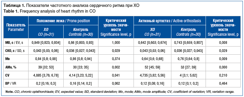

The study analyzed vegetative changes in the groups through frequency and spectral analysis of heart rhythm parameters. The evaluations were conducted both at rest (in supine position) and in active orthostasis.
To study the functions of the ANS, the Baevsky method of mathematical analysis of heart rhythm was used, which provides a comprehensive illustration of the organism's adaptation and compensatory mechanisms [12]. The following parameters characterizing the activity of different ANS branches were calculated:
-
E is the expected value (or mathematical expectation) of the dynamic series of cardio-intervals used to reflect the final result of all regulatory influences on the heart and circulatory system as a whole. E is one of the most stable mathematical and statistical parameters of the organism, as it is well homeostatizable. Deviations from individual normal values typically indicate an increase in the load on the circulatory system or the presence of pathological deviations;
-
RMS is the root-mean-square deviation of the values of the dynamic series of cardio-intervals and is one of the key parameters of HRV that characterizes the status of regulation mechanisms;
-
Mo (the mode, which represents the humoral channel of HR central regulation);
-
AMo (the amplitude of the mode, which represents the central nervous branch of
-
sympathetic regulation);
-
CV (the coefficient of variation, which is a normalized value of RMS);
-
R (the range of variability, which represents the autonomous parasympathetic branch) [12].
The parameters of HR spectral composition were also analyzed:
-
HF, or high-frequency respiratory waves, with an average power of the spectral function between 0.15-0.35 Hz/s2, characterize the activity of autonomous regulation, predominantly parasympathetic stimuli;
-
LF, or first-order low-frequency slow waves, with an average power of spectral functions between 0.05-0.15 Hz/s2, characterize the activity of central regulation, predominantly sympathetic stimuli;
-
VLF refers to second-order very low frequency slow waves, which are characterized by the impairment of humoral regulation.
Integral parameters (indices) characterizing the ratios of primary parameters were calcu-lated:
-
TI, the tension index that describes the degree of centralization of HR control; VBI, the vegetative balance index, which is the ratio of sympathetic and parasympathetic activity;
-
IARP, the indicator of adequacy of regulatory processes;
-
VRI, vegetative rhythm indicator;
-
OI, orthostatic index, measures changes in ANS function [12, 13].
The results were processed using the original CORVEG program (Russian Patent and Trademark Agency Certificate of Official Registration No. 200061883, dated September 8, 2000). Standard deviation of all normal RR intervals (SDNN) and the difference between the maximum hourly HRV (circadian rhythm) were calculated as standard measures of HRV over time. Spectral power was calculated using both Fast Fourier Transform and autoregressive model in four frequency ranges. VLF spectral components were calculated over the entire recording interval using the Fast Fourier Transform, specifically in the ranges of 0.003 to 0.04 Hz/s2 (VLF), 0.04 to 0.15 Hz/s2 (LF), and 0.15 to 0.40 Hz/s2 (HF). LF and HF components were calculated through autoregression of 100 RR intervals.
Statistical analysis was conducted using Statistica 8.0 software (StatSoft Inc.). Quantitative parameters were presented as median, upper and lower quartiles (Me [LQ; HQ]), while qualitative parameters were presented as frequencies (percentages). The Mann-Whitney U test was used to compare two groups of quantitative parameters. The study compared group frequencies using the Pearson χ2 test. In cases wh ere the number of observations was less than 5 in any cell of the contingency table, the Monte Carlo simulation was applied. Residuals and their significance were calculated to identify non-random contributions to the criterion statistics. The null hypothesis had a critical significance level of 0.05.
Results and Discussion
Tables 1 and 2 present the findings on autonomic changes in patients with CO and controls. In patients with CO, frequency analysis revealed significant deviations of RMS, AMo, and CV in the supine position and E, RMS, Mo, AMo in active orthostasis. This illustrates an increase in the effect of the sympathetic branch and a reduction in the effect of the parasympathetic branch of the ANS, resulting in a decrease in HRV in the CO group. In the supine position, HF and LF were significantly lower in the CO group (p<0.05), indicating impaired humoral regulation and decreased activity of intersystem regulation.
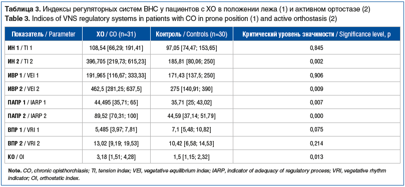
Based on the above-mentioned values, ANS regulatory system indices were calculated. The CO group had significantly higher TI in orthostasis than the control group (p<0.05), indicating a shift in autonomic balance towards the sympathetic system. Additionally, VBI in active orthostasis was significantly higher, indicating increased sympathetic regulation. The supine position and orthostasis resulted in significantly higher IARP values, indicating greater centralization of heart rhythm control in CO patients. The CO group also exhibited significantly higher OI values, indicating increased sympathetic influences (see Table 3).
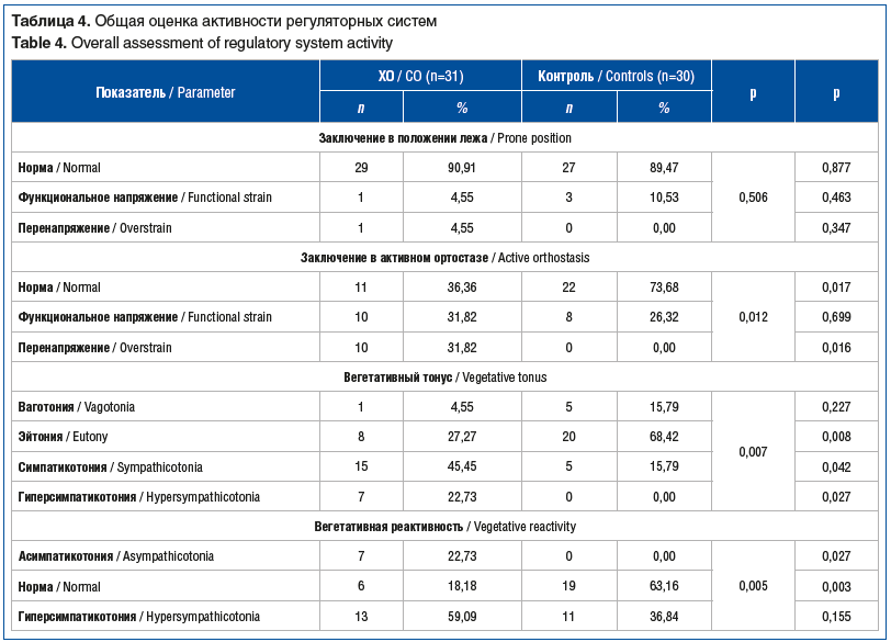
Changes in HR regulation parameters showed significantly increased sympathetic regulation at rest and orthostasis in CO patients (p<0.05). Moreover, the study found that in both resting and exercise states, the patients' autonomic homeostasis shifted towards the sympathetic nervous system. In 31% of patients, central dysregulation was detected. Additionally, autonomic tone and reactivity in CO were significantly shifted towards sympathetic and hypersympathetic tonus (see Table 4).
Optimal HRV indicates normal functioning and good adaptive abilities of the human body in various pathogenic conditions [29, 30]. Chronic stress can lead to decreased HRV and impaired physiological functioning and energy utilization [31, 32]. This may indicate age-related systemic depletion or pathological functioning of self-regulation and adaptation systems [27, 33, 34]. Frequency and spectral analysis of HRV in patients with cholangitis have revealed deviations at all levels of autonomic regulation. These deviations manifest as increased sympathetic regulation and decreased parasympathetic regulation, vasoreflex and baroreflex mechanisms, and humoral regulation. These disorders are caused not only by local damage to the biliary tract (inflammation of bile ducts) and the reflex effect of the gallbladder on HR, but also by the systemic toxic effect of opisthorchis invasion on the organism.
Conclusions
Therefore, patients with CO exhibit impaired functionality of the ANS and decreased HRV compared to the control group. Treatment approaches for CO should include tools that affect vegetative status and reactivity, improve neurohumoral homeostasis during de-worming and convalescence, and consider pharmacotherapy of ANS status as a necessary prerequisite for successful convalescence of CO patients. Vegetotrophic agents such as β-blockers, potassium, and magnesium preparations may be the preferred drugs.
About the authors:
Ekaterina Yu. Plotnikova — Dr. Sc. (Med.), professor of the Department of Training of Primary Care Doctors, Head of the Course of Clinical Gastroenterology, Kemerovo State Medical University; 22A, Voroshilov str., Kemerovo, 650029, Russian Federation; ORCID iD 0000-0002-6150-1808.
Nina V. Karbysheva — Dr. Sc. (Med.), Professor, Head of the Department of Infectious Diseases with the Course of Additional Professional Education, Altai State Medical University; 40, Lenin str., Barnaul, 656038, Russian Federation; ORCID iD 0000-0001-8320-3468.
Marina A. Nikonorova — Dr. Sc. (Med.), Associate Professor, professor of the Department of Infectious Diseases with the Course of Additional Professional Education, Altai State Medical University; 40, Lenin str., Barnaul, 656038, Russian Federation; ORCID iD 0000-0001-6621-9310.
Olga V. Beskhlebova — C. Sc. (Med.), Associate Professor, associate professor of the Department of Infectious Diseases with the Course of Additional Professional Education, Altai State Medical University; 40, Lenin str., Barnaul, 656038, Russian Federation; ORCID iD 0000-0003-4561-1019.
Mariya E. Ivanova — Head of the Second Infectious Department, Altay Kray Clinical Center for Maternity and Childhood; 179, Gushchin str., Barnaul, 656019, Russian Federation; ORCID iD 0009-0007-9413-3216.
Maksim A. Sinkov — C. Sc. (Med.), cardiovascular surgeon, Scientific Institute for Complex Issues of Cardiovascular Diseases; 6, Sosnovyy blvd., Kemerovo, 650002, Russian Federation; ORCID iD 0000-0002-2494-8694.
Ekaterina V. Kanner — Dr. Sc. (Med.), Senior Researcher of the Clinical Department of Infectious Pathology, Central Research Institute of Epidemiology of the Russian Federal Service for Supervision of Consumer Rights Protection and Human Well-Being; 3А, Novogireevskaya str., Moscow, 111123, Russian Federation; ORCID iD 0000-0003-3204-1846.
Ilya D. Kanner — 6th year student of Lomonosov Moscow State University; 1, Leninskie Gory, Moscow, 119991, Russian Federation; ORCID iD 0000-0003-4632-0547.
Contact information: Ekaterina Yu. Plotnikova, e-mail: eka-pl@rambler.ru.
Financial Disclosure: no authors have a financial or property interest in any material or method mentioned.
There is no conflict of interest.
Received 09.08.2023.
Revised 30.09.2023.
Accepted 20.10.2023.
2. Kiyan V.S., Bulashev A.K., Katokhin A.V. Opisthorchis felineus and Metorchis bilis Metacercariae in Cyprinid Fish Leuciscus idus in Nura-Sarysu River, Kazakhstan. Korean J Parasitol. 2018;56(3):267–274. DOI: 10.3347/kjp.2018.56.3.267.
3. Плотникова Е.Ю., Баранова Е.Н. Проблемы лечения описторхозной инвазии. РМЖ. Медицинское обозрение. 2018;3:53–56. [Plotnikova E.Yu., Baranova Е.N. Problems of treatment of opisthorchiasis. RMJ. Medical Review. 2018;3:53–56 (in Russ.)].
4. Pozio E., Gomez-Morales M.A. Clonorchiasis and opisthorchiasis. In: Helminth Infections and Their Impact on Global Public Health. Bruschi F., ed. Springer-Nature: Cham, Switzerland; 2022:221–256. DOI: 10.1007/978-3-031-00303-5.
5. Armignacco O., Caterini L., Marucci G. et al. Human illnesses caused by Opisthorchis felineus flukes, Italy. Emerg Infect Dis. 2008;14(12):1902–1905. DOI: 10.3201/eid1412.080782.
6. Saijuntha W., Sithithaworn P., Petney T.N., Andrews R.H. Foodborne zoonotic parasites of the family Opisthorchiidae. Res Vet Sci. 2021;135:404–411. DOI: 10.1016/j.rvsc.2020.10.024.
7. De Liberato C., Scaramozzino P., Brozzi A. et al. Investigation on Opisthorchis felineus occurrence and life cycle in Italy. Vet Parasitol. 2011;177(1-2):67–71. DOI: 10.1016/j.vetpar.2010.11.042.
8. Caffara M., Serracca L., Gustinelli A. et al. Development and validation of species-specific molecular diagnostic tool for Opisthorchis felineus (Digenea, Opisthorchiidae) metacercariae. Int J Food Microbiol. 2017;242:98–100. DOI: 10.1016/j.ijfoodmicro.2016.11.024.
9. Яблоков Д.Д. Описторхоз человека. Томск: Изд-во Том. ун-та; 1979. [Yablokov D.D. Human opisthorchiasis. Tomsk: Izd-vo Tom. un-ta; 1979 (in Russ.)].
10. О состоянии санитарно-эпидемиологического благополучия населения в Российской Федерации в 2020 году: Государственный доклад. М.: Федеральная служба по надзору в сфере защиты прав потребителей и благополучия человека, 2021. [On the state of sanitary and epidemiological well-being of the population in Russian Federation in 2020: State report. Federal'naya sluzhba po nadzoru v sfere zashchity prav potrebiteley I blagopoluchiya cheloveka. M.: 2021 (in Russ.)].
11. Cai Z., Cheng H., Xing Y. et al. Autonomic nervous activity analysis based on visibility graph complex networks and skin sympathetic nerve activity. Front Physiol. 2022:13:1001415. DOI: 10.3389/fphys.2022.1001415.
12. Баевский Р.М., Кириллов О.В., Клецкин С.З. Математический анализ сердечного ритма при стрессе. М.: Наука; 1984. [Bayevskiy R.M., Kirillov O.V., Kletskin S.Z. Mathematical analysis of heart rate under stress. M.: Nauka; 1984 (in Russ.)].
13. Zemaityte D.J., Varoneckas G.A., Sokolov E.N. Interaction between the parasympathetic and sympathetic divisions of the autonomic nervous system in cardiac rhythm regulation. Hum Physiol. 1985;11(3):208–215.
14. Heart rate variability. Standards of measurement, physiological interpretation, and clinical use. Task Force of the European Society of Cardiology and the North American Society of Pacing and Electrophysiology. Circulation. 1996;93:1043–1065. DOI: 10.1161/01.CIR.93.5.1043.
15. Thomas B.L., Claassen N., Becker P., Viljoen M. Validity of Commonly Used Heart Rate Variability Markers of Autonomic Nervous System Function. Neuropsychobiology. 2019;78:14–26. DOI: 10.1159/000495519.
16. Skorić K.M., Adamec I., Cifrek M., Habek M. Analysis of Autonomic Nervous System Biosignals. In: CMBEBIH 2021. Badnjevic A., Gurbeta Pokvić L., eds. Springer: Cham; 2021:20–27. DOI: 10.1007/978-3-030-73909-6_3.
17. McCraty R., Shaffer F. Heart rate variability: new perspectives on physiological mechanisms, assessment of self-regulatory capacity, and health risk. Glob Adv Health Med. 2015;4:46–61. DOI: 10.7453/gahmj.2014.073.
18. Grässler B., Dordevic M., Darius S. et al. Age-Related Differences in Cardiac Autonomic Control at Resting State and in Response to Mental Stress. Diagnostics (Basel). 2021;11(12):2218. DOI: 10.3390/diagnostics11122218.
19. Laborde S., Mosley E., Thayer J.F. Heart Rate Variability and Cardiac Vagal Tone in Psychophysiological Research — Recommendations for Experiment Planning, Data Analysis, and Data Reporting. Front Psychol. 2017;8:213. DOI: 10.3389/fpsyg.2017.00213.
20. Shaffer F., Ginsberg J.P. An Overview of Heart Rate Variability Metrics and Norms. Front Public Health. 2017;5:258. DOI: 10.3389/fpubh.2017.00258.
21. Lane R.D., Reiman E.M., Ahem G.L., Thayer J.F. Activity in medial prefrontal cortex correlates with vagal component of heart rate variability during emotion. Brain Cognit. 2001;47:97–100.
22. Goldberger A.L. Is the normal heartbeat chaotic or homeostatic? News Physiol Sci. 1991;6:87–91.
23. Рябыкина Г.В., Соболев А.В. Анализ вариабельности ритма сердца. Кардиология. 1996;36(10):87–97. [Ryabykina G.V., Sobolev A.V. Analysis of heart rate variability. Kardiologiya. 1996;36(10):87–97 (in Russ.)].
24. Beckers F., Verheyden B., Aubert A.E. Aging and nonlinear heart rate control in a healthy population. Am J Physiol Heart Circ Physiol. 2006;290(6):H2560–H2570. DOI: 10.1152/ajpheart.00903.2005.
25. Vaillancourt D.E., Newell K.M. Changing complexity in human behavior and physiology through aging and disease. Neurobiol Aging. 2002;23(1):1–11. DOI: 10.1016/S0197-4580(01)00247-0.
26. Peltola M.A. Role of Editing of R–R Intervals in the Analysis of Heart Rate Variability. Front Physiol. 2012;3:148. DOI: 10.3389/fphys.2012.00148.
27. Heart rate variability. Standards of measurement, physiological interpretation, and clinical use. Task Force of the European Society of Cardiology and the North American Society of Pacing and Electrophysiology. Eur Heart J. 1996;17(3):354–381.
28. Berntson G.G., Stowell J.R. ECG artifacts and heart period variability: don't miss a beat! Psychophysiology. 1998;35(1):127–132.
29. Singer D.H. High heart rate variability, marker of healthy longevity. Am J Cardiol. 2010;106(6):910. DOI: 10.1016/j.amjcard.2010.06.038.
30. McCraty R., Shaffer F. Heart Rate Variability: New Perspectives on Physiological Mechanisms, Assessment of Self-regulatory Capacity, and Health risk. Glob Adv Health Med. 2015;4(1):46–61. DOI: 10.7453/gahmj.2014.073.
31. Geisler F.C., Kubiak T., Siewert K., Weber H. Cardiac vagal tone is associated with social engagement and self-regulation. Biol Psychol. 2013;93(2):279–286. DOI: 10.1016/j.biopsycho.2013.02.013.
32. Reynard A., Gevirtz R., Berlow R. et al. Heart rate variability as a marker of self-regulation. Appl Psychophysiol Biofeedback. 2011;36(3):209–215. DOI: 10.1007/s10484-011-9162-1.
33. Singer D.H., Martin G.J., Magid N. et al. Low heart rate variability and sudden cardiac death. J Electrocardiol. 1988;21 Suppl:S46–S55. DOI: 10.1016/0022-0736(88)90055-6.
34. Thayer J.F., Hansen A.L., Saus-Rose E., Johnsen B.H. Heart rate variability, prefrontal neural function, and cognitive performance: the neurovisceral integration perspective on self-regulation, adaptation, and health. Ann Behav Med. 2009;37(2):141–153. DOI: 10.1007/s12160-009-9101-z.

This work is licensed under a Creative Commons «Attribution» 4.0 License.
