Gastrointestinal stromal tumor of the stomach (clinical case)




















DOI: 10.32364/2587-6821-2021-5-6-433-437
T.E. Skvortsova1, I.A. Oganezova1, O.I. Medvedeva1, O.M. Zhorina1, I.A. Belyakov2, S.L. Vorobiev2
1I.I. Mechnikov North-Western State Medical University, St. Petersburg, Russian Federation
2National Center for Clinical Morphological Diagnostics LLC, St. Petersburg, Russian Federation
Gastrointestinal stromal tumors (GIST) are the most common malignant submucosal neoplasms of the gastrointestinal tract, with predominant localization in the stomach and small intestine. The disease pattern is the absence of specific clinical signs, at least a third of cases are detected during examinations or surgical interventions for other diseases or at an autopsy. The examination algorithm of patients with suspected GIST includes endoscopic and radiation diagnostic methods, including a number of high-tech and expensive techniques, such as endoscopic ultrasonography, computed tomography, immunohistochemical and molecular genetic methods for studying biopsy material. Only conducting a full set of examinations will allow timely diagnosis and choosing the optimal treatment tactics for the disease with a high risk of malignancy. The authors present their own clinical case of a patient, female, with GIST, which is of considerable interest to clinicians. The above clinical case represents a typical clinical and morphological picture of GIST. The disease onset occurred in a patient over the age of 60 years and proceeded under the guise of organic dyspepsia. In this case, the use of expensive high-tech techniques allowed to verify the diagnosis of GIST and refer the patient to surgical treatment.
Keywords: gastrointestinal stromal tumor, stomach tumor, endoscopic ultrasonography, immunohistochemistry, differential diagnosis.
For citation: Skvortsova T.E., Oganezova I.A., Medvedeva O.I. et al. Gastrointestinal stromal tumor of the stomach (clinical case). Russian Medical Inquiry. 2021;5(6):433–437 (in Russ.). DOI: 10.32364/2587-6821-2021-5-6-433-437.
Background
Gastrointestinal stromal tumors (GISTs) are rare mesenchymal neoplasms, with an annual incidence of approximately 7 to 20 cases per one million. Meanwhile, GISTs are the most common malignancies among gastrointestinal (GI) submucosal tumors. GISTs are generally diagnosed in patients over 60 without gender preference. GISTs are found along the entire GI tract, most commonly in the stomach (50-60%) or small intestine (25-35%), colon (<7%) or rectum (<5%), and, rarely, esophagus (1%). Primary GISTs are sometimes localized outside hollow organs, e.g., in the omentum or retroperitoneal space [1, 2].
Histologically, GISTs display spindle cell (70%), epithelioid cell (20%), or mixed cell (10%) patterns. In addition, cellular and nuclear polymorphism, areas of sclerosis and hyalinosis (sclerosing subtype), and myxoid stroma are found. Palisade-like GISTs are sometimes identified in the small intestine. The hypercell pattern of GISTs is characterized by a high mitotic activity [3].
Previously, GISTs were believed to originate from GI stromal cells. However, it was demonstrated recently that GISTs develop from interstitial cells of Cajal (ICC) as a result of oncogenic mutations of the receptor tyrosine kinase (KIT) or platelet-derived growth factor receptor alpha (PDGFR α). KIT mutations are found in 56-92% of GISTs, and PDGFR α mutations are found in 5-10% of GISTs. Meanwhile, mutations of KIT and PDGFR α are mutually exclusive. KIT mutations are commonly localized in exons 11 (70%) or 9 (10%) and, occasionally, exons 13 (1%) or 17 (1%). PDGFR α mutations are localized in exons 18 (5%), 12 (1%), or 14 (<1%). GISTs with PDGFR α mutations commonly originate from the stomach (90-93%). Wild-type GISTs without KIT or PDGFR α mutations (12-15%) are graded based on succinate dehydrogenase (SDH) deficiency. SDH-deficient tumors include Carney triad and Carney-Stratakis syndrome. Non-SDH-deficient tumors include neurofibromatosis type 1 and GISTs with BRAF, KRAS, and PIK3CA mutations. Wild-type GISTs are believed to have a lower malignant potential [4-6].
Clinical presentations, diagnosis, and differential diagnosis of GISTs
A feature of GISTs is an asymptomatic course at early stages and no specific clinical signs. According to published data, 15% to 30% of GISTs are asymptomatic and diagnosed accidentally, e.g., during examinations or surgical interventions for other diseases or autopsy (10%). As a tumor grows, non-specific symptoms (epigastric pain, fatigue, nausea, anorexia, weight loss, etc.) appear. Mucosal ulceration results in GI bleeding and anemia, requiring careful differential diagnosis with peptic gastric and duodenal ulcers. In addition, symptoms provoked by the compression/stenosis of a part of the GI tract (e.g., biliary obstruction in GIST of the Vater's ampulla, dysphagia in esophageal GIST, constipation in rectal GIST) are reported [7, 8].
The diagnostic algorithm for suspected GISTSs involves endoscopic and radiological tests. Endoscopic presentations are variable; tumor size varies from 1-2 cm to 20-30 cm. In small lesions, endoscopic presentations depend on growth patterns and include normal appearance (extra-organ growth), external compression (mixed growth), or submucosal masses (endo-organ growth). Differential diagnoses involve numerous nosologies, e.g., extra-organ compression, submucosal varicose veins, aberrant pancreas, infiltrative cancer, leiomyoma, schwannoma, lipoma, etc. Collecting an adequate histological sample to diagnose GIST during standard endoscopy is challenging since a tumor is covered by healthy mucosa [9-11].
Endoscopic ultrasound (EUS) is the crucial test for differential diagnosis that provides a precise distinguishing between GISTs (hypoechoic solid masses) and other submucosal lesions. In addition, EUS-assisted fine needle biopsy allows for collecting a sample of the submucosal tumor to perform histology and immunohistochemistry (IHC) [12, 13].
Computed tomography (CT) is used to clarify the diagnosis since this technique provides a clear view of tumor size and growth pattern, invasion of adjacent organs and tissues, and metastases. GISTs appear as lesions with a heterogeneous internal structure and sharp borders on CT scans. In addition, a significant irregular early "enhancement" of tumor masses is seen on contrast-enhanced CT scans [14, 15].
The primary technique to verify GIST is IHC that identifies specific markers. Expression of CD117 (KIT, up to 95% of GISTs), anoctamin 1 (DOG1, 87% of GISTs), CD34 (60-70% of GISTs), and other markers (smooth muscle actin, desmin, S-100, keratin) is typical for GISTs. According to the recommendations of expert oncological societies, GIST is verified by histological findings and IHC of biopsy samples (to identify these markers) [7, 11, 16–18].
Treatment and prognosis
Surgical intervention is now the critical treatment for localized and locally advanced GISTs. In unresectable tumors, metastases, or recurrent GISTs, tyrosine kinase inhibitors (imatinib) are prescribed [17, 19]. 10% to 30% of GISTs are potentially malignant. GISTs are graded based on malignancy risk, i.e., very low, low, moderate, or high. According to international consensuses, major prognostic factors for GISTs are mitotic index (mitotic count in a 5-mm2 area of tumor at 400× magnification), tumor size, and localization. The risk of metastasis increases depending on the size of the primary tumor but irrespective of the mitotic index. Ki-67, a marker of proliferative activity, is also important. Its level significantly increases as the risk of GIST aggressiveness increases. Ki-67 is used to evaluate malignancy and prognosis. It is generally accepted that the outcome of gastric GISTs (if other characteristics are equal) is more favorable [18, 20, 21].
Mutations in various exons of KIT and PDGFR α are relevant prognostic markers indicative of clinical course and expected response to treatment with tyrosine kinase inhibitors [22]. A meta-analysis by L. Yan et al. [23] which included 20 studies and 3,780 patients has demonstrated that progression-free survival in GISTs is significantly lower in KIT exon 9 mutations than in KIT exon 11 mutations (odds ratio/OR 3.60, 95% confidence interval/CI 2.17-5.98, z=4.96, p<0.00001, heterogeneity I2= 0%). KIT mutations are more common in patients with large (≥5 cm) GISTs (OR 1.51, 95% CI 1.05-2.17, z=2.22, p=0.03, heterogeneity I2=0%, p=0.97) and higher (≥5/50 HPFs) mitotic index (OR 1.76, 95% CI 1.05-2.95, z=2.15, p=0.03, heterogeneity I2=57%, p=003).
Case report
A 67-year-old woman was referred to a general practitioner in March 2019 with complaints of mild epigastric pain, dizziness associated with pain, nausea after eating, heartburn, and hypogastric pain not related to defecation or urination. Daily bowel movements, Bristol type 4, no pathological admixtures. The patient felt worse for two months. She suffered from chronic gastritis and cholelithiasis for a long time. Physical examination: the patient is in satisfactory condition, no cardiovascular and respiratory pathological signs, no abdominal distension, the abdomen is soft, abdominal pain on palpation of the epigastrium, and projection of the transverse colon. CBC parameters are within normal ranges.
Abdominal ultrasound (April 08, 2019)demonstrated no liver enlargement, normal hepatic echogenicity. The diameter of the portal vein is 10 mm. The diameter of the choledoch is 4 mm, kinking of the gallbladder, wall thickness is 2 mm, echo positive mobile sludge. The pancreas is sized 16×10×14 mm, hypoechoic lesion in the body sized 21×20 mm.
To specify pathological signs, chest and abdominal contrast (iopamidol)-enhanced CT was performed on April 23, 2019. Chest CT identified enhanced bilateral lesions with sharp, irregular, lumpy borders with the increase in density from 13 HU to 93 HU (lesions in the S3 segment sized 8×10 mm and in the axillary subsegment sized up to 7 mm in the right lung, and lesion in the S5 segment sized up to 3 mm), small consolidations in the right S9 segment and left S6 segment. No enlargement of intrathoracic lymph nodes.
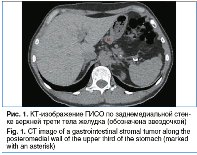
Abdominal CT identified an enhanced lesion outside the posterior medial wall of the upper third of the stomach sized 18×21×13 mm, with sharp borders with the increase in density from 41 HU to 71 HU (see Fig. 1). The tumor lies adjacent to the stomach along 10 mm. Homogeneous parenchymal enhancement of the liver. No enlargement of intra- and extrahepatic biliary ducts. No enlargement of the gallbladder, homogeneous content, no concrements, no wall thickening. No enlargement of the portal and splenic veins. No enlargement of the spleen; its structure is homogeneous. No enlargement of the pancreas, its structure is homogeneous, no contrast-enhanced lesions, no infiltration of the peripancreatic fat. Degenerative lesions of the thoracic and lumbar spine. The heterogeneous structure of the thoracic and lumbar vertebrae and iliac bones results from multiple rarefied areas with sharp borders. Areas of osteosclerosis in the L4 body sized 4 mm and left iliac bone sized 4×6 mm. Hemangioma of the T9 body.
To specify bone abnormalities, bone scintigraphy was performed that provided no strong evidence of specific skeletal lesions.
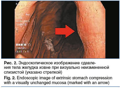
Given the exophytic tumor originating from the stomach, endoscopy was performed on April 30, 2019. The esophagus is patent, hyperemic mucosa, caseous whitish fixed deposit. A cardiac rosette is opened. Gastric mucosa prolapses to the esophagus. A Schatzki ring with vulnerable mucosa is seen at the gastroesophageal junction. The typical shape of the stomach. Diffuse mucosal hyperemia, four acute erosions covered with fibrin sized up to 3 mm, are found in the antrum. Mild narrowing of the stomach lumen by the extrinsic compression of its posterior wall (mucosa is unaffected) is reported (see Fig. 2).
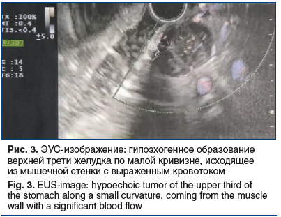
To specify tumor variant, EUS was performed on April 17, 2019. A rounded submucosal tumor sized up to 15 mm was found in the upper third of the lesser curvature of the stomach. The tumor is moderately prominent in the stomach lumen without its deformation and covered with unaffected mucosa. The tumor is dense under instrumental palpation. In addition, US scanning (7.5-12 MHz) of the gastric wall identified a rounded, non-homogeneous, predominantly hypoechoic tumor sized up to 20 mm originated from the muscular layer, with predominantly extra-organ growth and significant blood circulation (see Fig. 3).
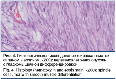
After an antifungal treatment course (fluconazole 150 mg for 14 days), the patient underwent video-assisted endoscopic enucleation of an extra-organ tumor of the stomach body (July 10, 2019). Histologically, this rounded, encapsulated, mesenchymal tumor sized 2.7 cm is composed of spindle cells with abundant collagen matrix and no cell polymorphism. This pattern is typical for sclerosing GIST. Additional histology was performed in the National Center of Clinical Morphological Diagnostics. Macroscopically, the excised tumor represents a rounded, dense node sized 2.7 cm, with a granular surface, grey and fibrous on the cut surface, with dot hemorrhages. Microscopically, this well-circumscribed mesenchymal tumor sized 2.7 cm is composed of spindle cells with smooth cell differentiation, minimal cellular nuclear polymorphism, and areas of abundant collagenization. This pattern is typical for sclerosing GIST (see Fig. 4).
IHC revealed fragments of a spindle-cell tumor with the areas of a moiré pattern, focal fibrosis, and hyalinosis. Mitotic activity is low (less than 5 mitoses in a 5-mm2 area of tumor at 400× magnification). Tumor cells express CD34 and DOG1 (diffusely) and desmin (focally) but do not express S-100 (see Fig. 5). Ki-67 is 2-5%.
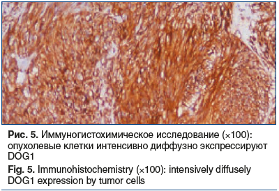
Impression: histological pattern and immune phenotype of the tumor correspond to GIST group 2.
The woman was scheduled for surgical intervention.
Conclusions
GISTs are rare mesenchymal submucosal GI malignancies without characteristic symptoms whose diagnosis is challenging. As a result, 50% of these tumors have metastases by the time of diagnosis. Management strategy involves radical surgery of localized and locally advanced GISTs with a 5-year survival rate of 54-65% [2, 19].
About the authors:
Tatyana E. Skvortsova — C. Sc. (Med.), Associate Professor, Associate Professor of the Department of Propaedeutics of Internal Diseases, Gastroenterology and Nutrition named after S.M. Ryss, I.I. Mechnikov North-Western State Medical University; 41, Kirochnaya str., St. Petersburg, 191015, Russian Federation; ORCID iD 0000-0002-9362-9961.
Inna A. Oganezova — Dr. Sc. (Med.), Professor, Professor of the Department of Propaedeutics of Internal Diseases, Gastroenterology and Nutrition named after S.M. Ryss, I.I. Mechnikov North-Western State Medical University; 41, Kirochnaya str., St. Petersburg, 191015, Russian Federation; ORCID iD 0000-0003-0844-4469.
Olga I. Medvedeva — C. Sc. (Med.), Associate Professor, Associate Professor of the Department of Propaedeutics of Internal Diseases, Gastroenterology and Nutrition named after S.M. Ryss, I.I. Mechnikov North-Western State Medical University; 41, Kirochnaya str., St. Petersburg, 191015, Russian Federation; ORCID iD 0000-0002-9730-7361.
Olga M. Zhorina — C. Sc. (Med.), Head of the Department of Computed Tomography at the Clinic named after Peter the Great, Associate Professor of the Department of Radiation Diagnostics and Therapy, I.I. Mechnikov North-Western State Medical University; 41, Kirochnaya str., St. Petersburg, 191015, Russian Federation; ORCID iD 0000-0002-9116-737X.
Ilya A. Belyakov — Head of the Department of Autopsy, National Center for Clinical Morphological Diagnostics LLC; 8 bld. 2A, O. Dundich str., St. Petersburg, 192283, Russian Federation; ORCID iD 0000-0002-9100-7608.
Sergey L. Vorobiev — C. Sc., Director, National Center for Clinical Morphological Diagnostics LLC; 8 bld. 2A, O. Dundich str., St. Petersburg, 192283, Russian Federation; ORCID iD 000-0002-7817-9069.
Contact information: Inna A. Oganezova, e-mail: oganezova@rambler.ru.
Financial Disclosure: no authors have a financial or property interest in any material or method mentioned.
There is no conflict of interests.
Received 14.07.2021.
Revised 06.08.2021.
Accepted 31.08.2021.
2. Вашакамадзе Л.А., Колобаев И.В., Беспалов П.Д. и др. Гастроинтестинальные стромальные опухоли: классификация, диагностика и лечение. Онкология. Журнал им. П.А. Герцена. 2013;2(2):66–70. [Vashakamadze L.A., Kolobaev I.V., Bespalov P.D. et al. Gastrointestinal stromal tumors: classification, diagnosis and treatment. Onkologiya Zhurnal im P.A. Gercena. 2013;2(2):66–70 (in Russ.)].
3. Халиков Д.Д., Ахметзянов Ф.Ш., Петров С.В. Клинико-морфологическая характеристика гастроинтестинальных стромальных опухолей. Архив патологии. 2017;79(4):48–55. [Khalikov D.D., Axmetzyanov F.Sh., Petrov S.V. Clinical and morphological characteristics of gastrointestinal stromal tumors. Arhiv patologii. 2017;79(4):48–55 (in Russ.)]. DOI: 10.17116/patol201779448-55.
4. Schaefer I.M., Mariño-Enríquez A., Fletcher J.A. What is New in Gastrointestinal Stromal Tumor? Adv Anat Pathol. 2017;24(5):259–267. DOI: 10.1097/PAP.0000000000000158.
5. Brcic I., Kashofer K., Skone D., Liegl-Atzwanger B. KIT mutation in a naïve succinate dehydrogenase-deficient gastric GIST. Genes Chromosomes Cancer. 2019;58(11):798–803. DOI: 10.1002/gcc.22768.
6. Liu Q.Y., Kong L.F., Xu Z.G. et al. Mutation of the KIT Gene, excluding Exon 11, in Gastrointestinal Stromal Tumors. Biomed Environ Sci. 2020;33(5):369–373. DOI: 10.3967/bes2020.050.
7. Бредихина Е.В., Бредихин Е.М. Диагностика и лечение гастроинтестинальных стромальных опухолей (обзор литературы). Медицинские новости. 2017;2:4–7. [Bredixina E.V., Bredixin E.M. Diagnostics and treatment of gastrointestinal stromal tumors (literature review). Medicinskie novosti. 2017;2:4–7 (in Russ.)].
8. Harris P.S., Romano J., Russ K.B. et al. Gastrointestinal Stromal Tumor: GIST Another Duodenal Ulcer. Ochsner J. 2020;20(2):236–238. DOI: 10.31486/toj.18.0167.
9. Cameron S., Beham A., Schildhaus H.U. Current Standard and Future Perspectives in the Treatment of Gastrointestinal Stromal Tumors. Digestion. 2017;95(4):262–268. DOI: 10.1159/000455835.
10. Nakano A., Nakamura M., Watanabe O. et al. Endoscopic Characteristics, Risk Grade, and Prognostic Prediction in Gastrointestinal Stromal Tumors of the Small Bowel. Digestion. 2017;95(2):122–131. DOI: 10.1159/000454728.
11. Akahoshi K., Oya M., Koga T., Shiratsuchi Y. Current clinical management of gastrointestinal stromal tumor. World J Gastroenterol. 2018;24(26):2806–2817. DOI: 10.3748/wjg.v24.i26.2806.
12. Khan S., Zhang R., Fang W. et al. Reliability of Endoscopic Ultrasound Using Miniprobes and Grayscale Histogram Analysis in Diagnosing Upper Gastrointestinal Subepithelial Lesions. Gastroenterol Res Pract. 2020;2020:6591341. DOI: 10.1155/2020/6591341.
13. Lee M.W., Kim G.H. Diagnosing Gastric Mesenchymal Tumors by Digital Endoscopic Ultrasonography Image Analysis. Clin Endosc. 2020;10.5946/ce.2020.061. DOI: 10.5946/ce.2020.061.
14. Jung H., Lee S.M., Kim Y.C. et al. A pictorial review on clinicopathologic and radiologic features of duodenal gastrointestinal stromal tumors. Diagn Interv Radiol. 2020;10.5152/dir.2019.19432. DOI: 10.5152/dir.2019.19432.
15. Peng F., Liu Y. Gastrointestinal Stromal Tumors of the Small Intestine: Progress in Diagnosis and Treatment Research. Cancer Manag Res. 2020;12:3877–3889. DOI: 10.2147/CMAR.S238227.
16. Nishida T., Blay J.Y., Hirota S. et al. The standard diagnosis, treatment, and follow-up of gastrointestinal stromal tumors based on guidelines. Gastric Cancer. 2016;19(1):3–14. DOI: 10.1007/s10120-015-0526-8.
17. Casali P.G., Abecassis N., Bauer S. et al. Gastrointestinal stromal tumours: ESMO-EURACAN Clinical. Practice Guidelines for diagnosis, treatment and follow-up. Ann Oncol. 2018;29:iv68–78. DOI: 10.1093/annonc/mdy095.
18. Wu C.E., Tzen C.Y., Wang S.Y., Yeh C.N. Clinical Diagnosis of Gastrointestinal Stromal Tumor (GIST): From the Molecular Genetic Point of View. Cancers (Basel). 2019;11(5):679. DOI: 10.3390/cancers11050679.
19. Ahmed M. Recent advances in the management of gastrointestinal stromal tumor. World J Clin Cases. 2020;8(15):3142–3155. DOI: 10.12998/wjcc.v8.i15.3142.
20. Никулин М.П., Архири П.П., Владимирова Л.Ю. и др. Практические рекомендации по лекарственному лечению гастроинтестинальных стромальных опухолей. Злокачественные опухоли. 2020;10(3s2–1):510–518. [Nikulin M.P., Arxiri P.P., Vladimirova L.Yu. et al. Practical recommendations for the drug treatment of gastrointestinal stromalene tumors. Zlokachestvenny`e opuholi. 2020;10(3s2–1):510–518 (in Russ.)]. DOI: 10.18027/2224-5057-2020-10-3s2-29.
21. Zhang H., Liu Q. Prognostic Indicators for Gastrointestinal Stromal Tumors: A Review. Transl Oncol. 2020;13(10):100812. DOI: 10.1016/j.tranon.2020.100812.
22. Capelli L., Petracci E., Quagliuolo V. et al. Gastric GISTs: Analysis of c-Kit, PDGFRA and BRAF mutations in relation to prognosis and clinical pathological characteristics of patients — A GIRCG study. Eur J Surg Oncol. 2016;42(8):1206–1214. DOI: 10.1016/j.ejso.2016.05.022.
23. Yan L., Zou L., Zhao W. et al. Clinicopathological significance of c-KIT mutation in gastrointestinal stromal tumors: a systematic review and meta-analysis. Sci Rep. 2015;5:13718. DOI: 10.1038/srep13718.

This work is licensed under a Creative Commons «Attribution» 4.0 License.
