Non-invasive diagnostics of liver fibrosis: recent data on the possibilities of instrumental techniques




















DOI: 10.32364/2587-6821-2020-4-5-297-301
E.A. Kulebina, A.N. Surkov, O.V. Usoltseva
National Medical Research Center for Children’s Health, Moscow, Russian Federation
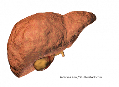
Liver fibrosis is a progressive pathological process that develops in chronic liver disease. This pathology can lead to cirrhosis. Liver biopsy remains the gold standard for evaluation of liver fibrosis, currently, though, non-invasive diagnostic methods are being actively introduced into clinical practice. Unlike liver biopsies, most non-invasive instrumental techniques are easy to perform, accessible, and repeatable, making them more convenient for evaluating dynamic fibrotic change. The review summarizes the main principles of non-invasive instrumental techinques for assessment of liver fibrosis severity, which have been studied and put into practice in recent years. The most widely used techniques in clinical practice are based on principles of elastography — it indirectly determines the organ tissue density. The techniques are characterized by different sensitivity and specificity in the diagnosis of fibrosis. The main issues arise when differentiating the disease stag es. For some techniques, the limitation of use is ascites, excessive subcutaneous fat, whereas the main limiting factor for others is the high cost of the procedure. Meanwhile, there is every reason to believe that non-invasive techniques can take the main place in the diagnosis of liver fibrosis and replace biopsy in the foreseeable future.
Keywords: liver fibrosis, chronic liver diseases, non-invasive diagnostic techniques, elastography, liver biopsy, magnetic resonance imaging, ultrasound.
For citation: Kulebina E.A., Surkov A.N., Usoltseva O.V. Non-invasive diagnostics of liver fibrosis: recent data on the possibilities of instrumental techniques. Russian Medical Inquiry. 2020;4(5):297–301. DOI: 10.32364/2587-6821-2020-4-5-297-301.
About the authors:
Elena A. Kulebina — pediatrician, gastroenterologist, potsgraduate student of Department of Gastroenterology, National Medical Research Center for Children's Health, 2, bld. 1, Lomonosovsky prospect, Moscow, 119296, Russian Federation, ORCID iD 0000-0001-9798-9617.
Andrey N. Surkov — Dr. of Sci. (Med), pediatrician, gastroenterologist, Head of Department of Gastroenterology with Hepatology Division, National Medical Research Center for Children's Health, 2, bld. 1, Lomonosovsky prospect, Moscow, 119296, Russian Federation, ORCID iD 0000-0002-3697-4283.
Olga V. Usoltseva — postgraduate student of Department of Gastroenterology with Hepatology Division, National Medical Research Center for Children's Health: 2, bld. 1, Lomonosovsky prospect, Moscow, 119296, Russian Federation, ORCID iD 0000-0003-0661-6976.
Contact information: Elena A. Kulebina, e-mail: e.kulebina@gmail.com Financial Disclosure: no authors have a financial or property interest in any material or method mentioned. There is no conflict of interests. Received 28.04.2020, revised 12.05.2020, accepted 26.05.2020.
2. Poynard T., Lenaour G., Vaillant J.C. et al. Liver biopsy analysis has a low level of performance for diagnosis of intermediate stages of fibrosis. Clin Gastroenterol Hepatol. 2012;10(6):657–663 e7. DOI: 10.1016/j.cgh.2012.01.023.
3. Castera L. Noninvasive assessment of liver fibrosis. Dig Dis. 2015;33(4):498–503. DOI: 10.1159/000374097.
4. Yin M., Venkatesh S.K. Ultrasound or MR elastography of liver: which one shall I use? Abdom Radiol (NY). 2018;43(7):1546–1551. DOI: 10.1007/s00261-017-1340-z.
5. Дворяковская Г.М., Ивлева С.А., Дворяковский И.В. и др. Комплексная ультразвуковая диагностика в оценке паренхимы печени и стадий фиброза у детей с аутоиммунным гепатитом. Российский педиатрический журнал. 2016;19(1):4–8. [Dvoryakovskaya G.M., Ivleva S.A., Dvoryakovskiy I.V. et al. Comprehensive ultrasound diagnosis in the evaluation of liver parenchyma and stages of fibrosis in children with autoimmune hepatitis Russian Pediatric J. 2016;19(1):4–8 (in Russ.)]. DOI 10.18821/1560-9561-2016-19-1-4-8.
6. Faria S.C., Ganesan K., Mwangi I. et al. MR imaging of liver fibrosis: current state of the art. Radiographics. 2009;29:1615–1635. DOI: 10.1148/rg.296095512.
7. Huwart L., Sempoux C., Vicaut E. et al. Magnetic resonance elastography for the noninvasive staging of liver fibrosis. Gastroenterology. 2008;135:32–40. DOI: 10.1053/j.gastro.2008.03.076.
8. Singh S., Venkatesh S.K., Wang Z. et al. Diagnostic performance of magnetic resonance elastography in staging liver fibrosis: a systematic review and meta-analysis of individual participant data. Clin Gastroenterol Hepatol. 2015;13:440–451. DOI: 10.1016/j.cgh.2014.09.046.
9. Loomba R., Wolfson T., Ang B. et al. Magnetic resonance elastography predicts advanced fibrosis in patients with nonalcoholic fatty liver disease: a prospective study. Hepatology. 2014;60:1920–1928. DOI: 10.1002/hep.27362.
10. Venkatesh S.K., Yin M., Takahashi N. et al. Non-invasive detection of liver fibrosis: MR imaging features vs. MR elastography. Abdom Imaging. 2015;40:766–775. DOI: 10.1007/s00261-015-0347-6.
11. Batheja M., Vargas H., Silva A.M. et al. Magnetic resonance elastography (MRE) in assessing hepatic fibrosis: performance in a cohort of patients with histological data. Abdom Imaging. 2015;40:760–765. DOI: 10.1007/s00261-014-0321-8.
12. Razek A.A., Abdalla A., Omran E. et al. Diagnosis and quantification of hepatic fibrosis in children with diffusion weighted MR imaging. Eur J Radiol. 2011;78:129–134. DOI: 10.1016/j.ejrad.2009.10.012.
13. Пеняева Э.И., Камалов Ю.Р., Сенча А.Н. и др. Ультразвуковое исследование с контрастным усилением в дифференциальной диагностике опухолевых образований печени. Медицинская визуализация. 2017;21(2):36–52. [Peniaeva E.I., Kamalov J.R., Sencha A.N. et al. Value of Contrast-Enhanced Ultrasound in Differential Diagnosis of Focal Live Lesions. Medical visualization. 2017;21(2):36–52 (in Russ)]. DOI: 10.24835/1607-0763-2017-2-36-52.
14. Abel M., Teoh W.C., Leen E. Contrast evaluation of liver masses. Ultrasound Clin. 2014;9(4):605–623.
15. Staub F., Tournoux-Facon C., Roumy J. et al. Liver fibrosis staging with contrast-enhanced ultrasonography: prospective multicenter study compared with METAVIR scoring. Eur Radiol. 2009;19:1991–1997. DOI: 10.1007/s00330-009-1313-x.
16. Gennisson J.L., Deffieux T., Fink M., Tanter M. Ultrasound elastography: principles and techniques. Diagn Interv Imaging. 2013;94:487–495. DOI: 10.1016/j.diii.2013.01.022.
17. Bamber J., Cosgrove D., Dietrich C.F. et al. EFSUMB guidelines and recommendations on the clinical use of ultrasound elastography. Part 1: Basic principles and technology. Ultraschall Med. 2013;34:169–184. DOI: 10.1055/s-0033-1335205.
18. Дворяковский И.В., Ивлева С.А., Дворяковская Г.М. и др. Cовременные технологии ультразвуковой диагностики цирроза при хронических болезнях печени у детей. Российский педиатрический журнал. 2016;19(4):202–208. [Dvoryakovskiy I.V., Ivleva S.A., Dvoryakovskaya G.M. et al. Modern technologies of ultrasound diagnostics of cirrhosis in chronic liver diseases in children. Russian Pediatric J. 2016;19(4):202–208 (in Russ.)]. DOI: 10.18821/1560-9561-2016-19-4-202-208.
19. Sandrin L., Fourquet B., Hasquenoph J.M. Transient elastography: a new noninvasive method for assessment of hepatic fibrosis. Ultrasound Med Biol. 2003;29(12):1705–1713. DOI: 10.1016/j.ultrasmedbio.2003.07.001.
20. Geng-lin Zhang, Qi-yi Zhao, Chao-shuang Lin et al. Transient Elastography and Ultrasonography: Optimal Evaluation of Liver Fibrosis and Cirrhosis in Patients with Chronic Hepatitis B Concurrent with Nonalcoholic Fatty Liver Disease. Biomed Res Int. 2019;2019:3951574. DOI: 10.1155/2019/3951574.
21. Chon Y.E., Choi E.H., Song K.J. et al. Performance of transient elastography for the staging of liver fibrosis in patients with chronic hepatitis B: a meta-analysis. PLoS One. 2012;7:e44930. DOI: 10.1371/journal.pone.0044930.
22. Tsochatzis E., Gurusamy K., Ntaoula S. et al. Elastography for the diagnosis of severity of fibrosis in chronic liver disease: a meta-analysis of diagnostic accuracy. J Hepatol. 2011;54:650–659. DOI: 10.1016/j.jhep.2010.07.033.
23. Cosgrove D., Piscaglia F., Bamber J. et al. EFSUMB guidelines and recommendations on the clinical use of ultrasound elastography. Part 2: Clinical applications. Ultraschall Med. 2013;34:238–253. DOI: 10.1055/s-0033-1335375.
24. European Association for the Study of the Liver. EASL Clinical Practice Guidelines: management of hepatitis C virus infection. J Hepatol. 2018;69(2):461–511. DOI: 10.1016/j.jhep.2018.03.026.
25. Poynard T., Munteanu M., Luckina E. et al. Liver fibrosis evaluation using real-time shear wave elastography: applicability and diagnostic performance using methods without a gold standard. J Hepatol. 2013;58:928–935. DOI: 10.1016/j.jhep.2012.12.021.
26. Bavu E., Gennisson J.L., Couade M. et al. Noninvasive in vivo liver fibrosis evaluation using supersonic shear imaging: a clinical study on 113 hepatitis C virus patients. Ultrasound Med Biol. 2011;37:1361–1373. DOI: 10.1016/j.ultrasmedbio.2011.05.016.
27. Ferraioli G., Tinelli C., Dal Bello B. et al. Accuracy of real-time shear wave elastography for assessing liver fibrosis in chronic hepatitis C: a pilot study. Hepatology. 2012;56:2125–2133. DOI: 10.1002/hep.25936.
28. Leung V., Shen J., Wong V. et al. Quantitative elastography of liver fibrosis and spleen stiffness in chronic hepatitis B carriers: comparison of shear-wave elastography and transient elastography with liver biopsy correlation. Radiology. 2013;269:910–918. DOI: 10.1148/radiol.13130128.
29. Zheng J., Guo H., Zeng J. et al. Two-dimensional shear-wave elastography and conventional US: the optimal evaluation of liver fibrosis and cirrhosis. Radiology. 2015;275:290–300. DOI: 10.1148/radiol.14140828.
30. Xiangdong Hu, Lanyan Qiu, Dong Liu, Linxue Qian. Acoustic Radiation Force Impulse (ARFI) Elastography for non‑invasive evaluation of hepatic fibrosis in chronic hepatitis B and C patients: a systematic review and meta-analysis. Med Ultrason. 2017;19(1):23–31. DOI: 10.11152/mu-942.
31. Sporea I., Sirli R.L., Deleanu A. et al. Acoustic radiation force impulse elastography as compared to transient elastography and liver biopsy in patients with chronic hepatopathies. Ultraschall Med. 2011;32(Suppl 1):S46–S52. DOI: 10.1055/s-0029-1245360.
32. Friedrich-Rust M., Nierhoff J., Lupsor M. et al. Performance of Acoustic Radiation Force Impulse imaging for the staging of liver fibrosis: a pooled meta-analysis. J Viral Hepat. 2012;19:e212–e219. DOI: 10.1111/j.1365-2893.2011.01537.x.
33. Sporea I., Bota S., Peck-Radosavljevic M. et al. Acoustic Radiation Force Impulse elastography for fibrosis evaluation in patients with chronic hepatitis C: an international multicenter study. Eur J Radiol. 2012;81:4112–4118. DOI: 10.1016/j.ejrad.2012.08.018.
34. Bota S., Herkner H., Sporea I. et al. Meta-analysis: ARFI elastography versus transient elastography for the evaluation of liver fibrosis. Liver Int. 2013;33:1138–1147. DOI: 10.1111/liv.12240.

This work is licensed under a Creative Commons «Attribution» 4.0 License.
