
Immediate outcomes of robot-assisted surgical interventions for gastric cancer
DOI: 10.32364/2587-6821-2023-7-6-3
S.A. Kovalenko1, D.V. Gladyshev1,2, M.E. Moiseev1, V.A. Vetoshkin1, V.Yu. Svitich1, S.S. Gnedash1
1St. Petersburg City Hospital No. 40, St. Petersburg, Russian Federation
2S.M. Kirov Military Medical Academy, St. Petersburg, Russian Federation
Background: stomach cancer is the 5th most common cancer worldwide and the 3rd leading cause of cancer death. In recent years, minimally invasive (including robot-assisted) techniques have been actively implemented in surgery for urological, gynecological, and gastrointestinal cancers.
Aim: to summarize the experience with robot-assisted surgical interventions for gastric cancer and to demonstrate direct surgical outcomes.
Patients and Methods: in 2018–2022, 111 patients (60 men and 51 women) with gastric cancer underwent robot-assisted surgery. The mean age was 64.6±5.5 years (33–78). The mean body mass index (BMI) was 27.9±2.8 kg/m2, 39.2% of patients had obesity (BMI>30 kg/m2). The tumor was located in the gastric cardia in 19 patients (17.1%), upper third of the stomach in 9 patients (8.1%), middle third of the stomach in 4 patients (3.6%), lower third of the stomach in 21 patients (18, 9%), and gastric antrum in 58 patients (52.3%). Distal subtotal gastric resection was performed in middle-third and lower-third gastric carcinomas. Gastrectomy was performed in the upper-third and gastric cardiac carcinoma. Two surgical systems, Da Vinci® Si and Da Vinci® Xi, were used for surgical interventions.
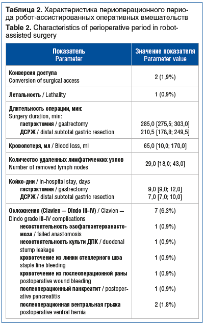
Results: 79 distal subtotal gastric resections and 32 gastrectomies were performed. The median duration of distal subtotal gastric resection and gastrectomy was 210.5 min [178.8; 249.5] and 285.0 min [275.5; 303.0], respectively. The median intraoperative blood loss was 65.0 ml [10.0; 170.0], and the median number of removed lymph nodes was 29.0 [18.0; 43.0]. Histological examination revealed clear resection margins (R0) in all patients. The conversion of surgical access was required in 2 patients (1.8%). Death was reported in 1 patient (0.9%). Clavien–Dindo grade III–IV postoperative complications were detected in 7 patients (6.3%). The median in-hospital stay was 7.0 days [7.0; 10.0] after distal subtotal gastric resection and 9.0 days [9.0; 12.0] after gastrectomy.
Conclusion: our findings support the global experience with robot-assisted surgical interventions for gastric cancer with minimal intra- and postoperative complications, which generally occur in elderly patients with severe comorbidities, obesity, and locally advanced cancer at baseline.
Keywords: robotic surgery, gastric cancer, oncology, gastric resection, gastrectomy.
For citation: Kovalenko S.A., Gladyshev D.V., Moiseev M.E., Vetoshkin V.A., Svitich V.Yu., Gnedash S.S. Immediate outcomes of robot-assisted surgical interventions for gastric cancer. Russian Medical Inquiry. 2023;7(6):353–358 (in Russ.). DOI: 10.32364/2587-6821-2023-7-6-3.
Background
Stomach cancer is the 5th most common cancer worldwide (13.5 per 100,000) and the 3rd leading cause of cancer death. According to the International Agency for Research on Cancer, 900,000 new diagnoses of stomach cancer and 650,000 deaths from this disease are reported annually worldwide. In the Russian Federation, stomach cancer ranks 3rdamong malignant disorders and occurs more often in men than in women (15.8% vs. 12.4%). Surgical outcomes (both independent and as part of a complex treatment) are still poor because of the lack of screening programs and referral at later stages in the setting of a latent disease course (in more than 60%, gastric cancer is detected at stage III-IV) [1]. This issue is also relevant in countries with a well-developed system of diagnosis and screening [2].
In recent years, minimally invasive (including robot-assisted) techniques have been actively implemented in surgery for urological, gynecological, and gastrointestinal (GI) cancers [3].
Despite more than 20 years of experience with robot-assisted technologies in gastric cancer surgery since its first use in 2001–2002 by M. Hashizume [4] and P.C. Giulianotti [5], this technique has not yet been included in any clinical guidelines. Several large meta-analyses (usually based on single-center non-randomized studies) have demonstrated the possibility, safety, and oncological adequacy of using a robotic complex for gastric cancer surgery [6]. The latest largest meta-analysis [7] included 48 retrospective studies, mainly from Asian countries (South Korea, Japan, China, Taiwan) and Italy, which compared laparoscopic and robot-assisted surgical interventions for gastric cancer of various locations (20,151 patients overall, 6,175 underwent robot-assisted gastrectomy and 13,976 underwent laparoscopic gastrectomy). The analysis yielded significant values (p<0.05). Currently, very little published data from large randomized clinical trials that comparatively assess robot-assisted technologies for gastric cancer surgery are available [8–10]. Thus, the authors [11] shared their experience with surgical interventions performed in 283 patients (141 robot-assisted distal gastrectomy and 142laparoscopic distal gastrectomy) and concluded that robot-assisted interventions for distal gastric cancer are associated with a lower rate of postoperative complications, less severe systemic inflammation, better quality of lymphadenectomy, shorter hospital stay, and faster recovery of GI tract functions.
According to the Analytical Department of Intuitive Surgical Inc. (2022), there are no more than 300 cases of robotic surgical complex gastric cancer surgery in the Russian Federation. Domestic publications on this issue are sporadic [12].
This study summarizes our experience with robot-assisted surgical interventions for gastric cancer and describes the immediate surgical outcomes.
Patients and Methods
In 2018–2022, 111 patients (60 men and 51 women) with gastric cancer aged 33–78 years underwent robot-assisted surgery. Of these, 39.2% had obesity (BMI>30 kg/m2).
All patients underwent examinations according to staging standards, including diagnostic laparoscopy with peritoneal washing cytology to identify circulating tumor cells. Somatic and nutritional status (based the NRS 2002 scale) and risks of surgery and anesthesia (based on the ASA physical status classification system) were evaluated. The Charlson comorbidity index (CCI) was used to assess comorbidities (see Table 1).
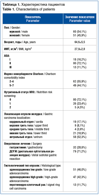
Poor nutritional status (5 patients) required additional preoperative treatment.
All patients underwent robot-assisted surgery using a technique described in detail earlier [13].
In tumors located in the middle and lower third of the stomach, distal subtotal gastrectomy (DSG) was performed. In tumors located in the upper third of the stomach and gastric cardia, gastrectomy was performed. In 5 patients (4.5%), DSG with Billroth 1 anastomosis was performed. In 74 patients (66.7%), DSG with Billroth 2 and Braun anastomosis was performed. In all patients who underwent gastrectomy, a linear-stapled Roux-en-Y gastric bypass was performed.
According to the preoperative instrumental tests, including staging laparoscopy, 7 patients (6.3%) were diagnosed with stage 1 gastric cancer, 60 patients (54.1%) with stage 2, 43 patients (38.7%) with stage 3, and 1 patient (0.9%) with stage 4. 42 patients (37.8%) underwent preoperative neoadjuvant FLOT (n=31) or mFOLFOX6 (n=11) chemotherapy. As a result, in some patients, significant tumor regression was reported. Distribution by stage (TNM classification of Malignant Tumors, 8th edition, 2017, UICC) based on postoperative histology was as follows: IA in 27 (24.3%), IB in 17 (15.3%), IIA in 19 (17.1%), IIB in 13 (11.7%), IIIA in 18 (16.2%), IIIB in 13 (11.7%), and IIIC in 4 (3.6%). Overall, 35 patients (31.5%) were diagnosed with stage III cancer. In 1 patient, the gastric tumor was staged as M1 because of the detection of atypical cells in the peritoneal washings. This patient underwent 4 cycles of FLOT chemotherapy. Control examination and restaging revealed significant tumor regression and no free tumor cells on repeated peritoneal washing cytology. This patient underwent surgical intervention (DSG), and surgical microscopic examination revealed complete regression. In 5 patients, tumor invasion into neighboring organs and tissues was reported (T4b): the tumor spread to the pancreatic capsule in 2 patients (1.8%), left liver lobe in 1 patient (0.9%), transverse mesocolon in 1 patient (0.9%), and crus of the diaphragm in 1 patient (0.9%).
Lymphadenectomy, Michel’s classification of hepatic artery variations, and ICG mapping. All patients underwent standard D2 lymphadenectomy [14] with the removal of 1, 3, 4sb, 4d, 5, 6, 7, 8a, 9, 11p, 12a groups of lymph nodes when performing DSG and 1, 2, 3, 4sa, 4sb. 4d, 5, 6, 7, 8a, 9, 11p, 11d, 12a groups of lymph nodes when performing total gastrectomy, except for 4 patients aged 78–80 years with a Charlson comorbidity index score of 6–7 who underwent D1+ lymphadenectomy. In 24 patients, standard D2 lymphadenectomy was supplemented with the removal of 8p and 12p groups of lymph nodes due to lymphadenopathy of these groups, as demonstrated by prehospital contrast-enhanced CT and intraoperatively. In 9 patients, the analysis of CT scans in the arterial phase revealed type II celiac trunk branching according to Michel’s classification, which was confirmed intraoperatively. This finding required isolation and preservation of the left hepatic artery and obligatory lymphadenectomy of the 7th and 9th groups of lymph nodes. In 1 patient, type IX branching according to Michel’s classification with the origination of the common hepatic artery from the superior mesenteric artery was detected. In 7 patients, augmented reality (AR) ICG angiography navigation identified contrast enhancement of group 14 lymph nodes, which required mobilization, removal, and marking. Further morphological examination of these lymph nodes revealed tumor growth in 2 patients to specify the disease stage and the amount of complex therapy.
Statistical analysis was performed using Excel 365 (Microsoft) and SPSS Statistics v. 26 (IBM). Quantitative data were summarized and represented as weighted arithmetic mean with a root mean square error (M±m) for normally distributed values and quartiles (Me [Q1; Q3]) for non-normally distributed values. Qualitative data were represented as absolute values and proportions (%). The Shapiro– Wilk test was applied to determine normality.
Results
Overall, 79 DSGs and 32 gastrectomies were performed. Extended surgical interventions were performed in 5 patients: resection of the transverse mesocolon (n=1), atypical resection of the S3 of the left liver lobe (n=1), resection of the right crus of the diaphragm (n=1), and resection of the pancreatic capsule (n=2). Conversion of the surgical access was required when performing DSG because of severe obesity (BMI 39.8 kg/m2) in one patient and tumor ingrowth into the pancreatic capsule and severe tumor infiltration along the splenic artery in another patient. The cause of the only death in the early postoperative period was an ischemic stroke after gastrectomy in a patient with severe systemic atherosclerosis and early encephalopathy.
The major characteristics of the perioperative period are listed in Table 2. Intraoperative blood transfusion was not required in any patient. Intraoperative complications (R. Rosenthal) grade 1 were reported in 3 patients and grade 2 in 1 patient, but they did not affect the surgical intervention course. Postoperatively, no patient required narcotic analgesics. Patient activation and sip feeding began on day 2 after surgery. Active intestinal peristalsis and passage of gas were reported on days 2-3.
Histology did not reveal positive surgical margins (R0). The tumor types comprised 24 Lauren intestinal type (21.7%), 58 Lauren diffuse type (52.2%), and 29 Lauren mixed type (32%) tumors.
The Clavien– Dindo classification was used to assess early postoperative complications. Clavien–Dindo grade III–IV postoperative complications were detected in 7 patients (6.3%) (see Table 2). Bleeding in the early postoperative period required re-laparoscopy and suturing. The failed esophagoenteric anastomosis was conservatively managed using intraluminal vacuum drainage. The failed duodenal stump was also managed conservatively (long-term drainage, formation of an external fistula, and antibacterial therapy). Postoperative pancreatitis occurred in a patient who underwent resection of the pancreatic capsule because of tumor invasion and was managed by prolonged drainage and anti-inflammatory therapy. Ventral incisional herniation after laparotomy during the late postoperative period required scheduled surgical treatment.
Among grade I–II complications, reflux gastritis (relieved after 6–9 months) was reported in 3 patients after the Billroth I procedure.
Learning curve
All robot-assisted surgical interventions described above were performed by three surgeons with many years of experience in open and laparoscopic surgeries for gastric cancer and open, laparoscopic, and robot-assisted surgeries for colorectal cancer. Indicators of reaching the “plateau” of the learning curve during the development and implementation of robot-assisted interventions for gastric cancer were a reduction in docking duration to 12–15 min and an overall duration of surgical intervention comparable to that of laparoscopic surgery (in case of standard anatomy and no tumor invasion into surrounding organs and tissues). These indicators were achieved after 10–15 procedures.
Discussion
Our clinical findings establish worldwide experience with robot-assisted surgical interventions for gastric cancer with a minimum of intra- and postoperative complications, which generally occur in elderly patients with severe comorbidities, obesity, and locally advanced tumors at baseline [15, 16]. Comparative analysis of our immediate results and foreign data demonstrates the similarity of findings, i.e., intraoperative blood loss, duration of surgical intervention, number of removed lymph nodes, and duration of hospital stay. In our study, the rate of postoperative complications was slightly lower (9% vs 12% in recent international meta-analysis). Meanwhile, a detailed analysis of grade III–V complications has shown that the relative number of anastomotic leaks, postoperative pancreatitis, and mortality is higher (1.8%, 0.9%, and 0.9%, respectively) than that in foreign studies (0.6%, 0.6%, and 0.4%, respectively). These differences are potentially accounted for by the relatively small sample size [7, 15]. In patients who underwent neoadjuvant chemotherapy during surgery, more severe fibrosis of the interfascial layers in the setting of a local inflammatory response was reported. This phenomenon compromised visualization and increased bleeding. However, no significant differences in the postoperative complication rate and quality of lymphadenectomy were reported in this group of patients. Robot-assisted surgical interventions help realize all the advantages of minimally invasive technologies, i.e., good cosmetic effect and mild pain that do not require postoperative narcotic analgesics. This contributed to early activation, lack of thromboembolism, early start of oral nutrition, and recovery of gastrointestinal function. Short recovery provides an earlier beginning of adjuvant drug therapy in some patients. Long-term oncological outcomes were not evaluated in this study. Meanwhile, given the volume of lymphadenectomy and R0 resections performed in 100%, one can suggest that robot-assisted surgical results will be comparable to open surgical results. The limitations of this study include the lack of comparison groups of laparoscopic and open surgical interventions for gastric cancer of various localizations and analysis of long-term oncological outcomes (5-year overall and relapse-free survival rates). The collection and retrospective analysis of the data are planned for publication.
Conclusions
Robot-assisted surgical interventions for gastric cancer are attractive because of their manufacturability. However, despite many years of global experience with this technique, no clear clinical guidelines are available for its use in certain categories of patients depending on localization, size, disease stage, and other characteristics. By sharing our clinical experience, we recommend robot-assisted surgery for localized gastric cancer stage T1–3 of any location without spreading to the esophagus and duodenum in patients of any gender and age with BMI of ≤30 kg/m2 and a risk of surgery and anesthesia of ≤ 3 (ASA physical status classification system). However, the technical advantages of this complex can also be realized in locally advanced tumors. Multicenter randomized clinical trials are required to obtain reliable data.
About the authors:
Sergey A. Kovalenko — oncologist of the Department of Abdominal Oncology, St. Petersburg City Hospital No. 40; 9, Borisova str., Sestroretsk, St. Petersburg, 197706, Russian Federation; ORCID iD 0000-0002-5850-0599.
Dmitry V. Gladyshev — Dr. Sc. (Med.), Deputy Chief Medical Officer of Surgery, St. Petersburg City Hospital No. 40; 9, Borisova str., Sestroretsk, St. Petersburg, 197706, Russian Federation; Associate Professor of the Department of Hospital Surgery of the S.M. Kirov Military Medical Academy; 6 Zh, Lebedeva str., St. Petersburg, 194044, Russian Federation; ORCID iD 0000-0001-5318-2619.
Mikhail E. Moiseev — oncologist of the Department of Abdominal Oncology, St. Petersburg City Hospital No. 40; 9, Borisova str., Sestroretsk, St. Petersburg, 197706, Russian Federation; ORCID iD 0000-0002-6180-6695.
Vyacheslav A. Vetoshkin — surgeon of the Department of Surgery for Anti-shock Measures, St. Petersburg St. Petersburg City Hospital No. 40; 9, Borisova str., Sestroretsk, St. Petersburg, 197706, Russian Federation; ORCID iD 0000- 0001-5346-9257.
Vadim Yu. Svitich — oncologist of the Department of Abdominal Oncology, St. Petersburg City Hospital No. 40; 9, Borisova str., Sestroretsk, St. Petersburg, 197706, Russian Federation; ORCID iD 0009-0002-2293-0527.
Sergey S. Gnedash — oncologist of the Department of Abdominal Oncology, St. Petersburg City Hospital No. 40; 9, Borisova str., Sestroretsk, St. Petersburg, 197706, Russian Federation; ORCID iD 0009-0003-4743-3014.
Contact information: Sergey A. Kovalenko, e-mail: kowalenko78@yandex.ru.
Financial Disclosure: no authors have a financial or property interest in any material or method mentioned.
There is no conflict of interest.
Received 27.02.2023.
Revised 23.03.2023.
Accepted 17.04.2023.
2. Van Boxel G.I., Ruurda J.P., van Hillegersberg R. Robotic-assisted gastrectomy for gastric cancer: a European perspective. Gastric Cancer. 2019;22(5):909–919. DOI: 10.1007/s10120-019-00979-z.
3. Bobo Z., Xin W., Jiang L. et al. Robotic gastrectomy versus laparoscopic gastrectomy for gastric cancer: meta-analysis and trial sequential analysis of prospective observational studies. Surg Endosc. 2019;33(4):1033–1048. DOI: 10.1007/s00464-018-06648-z.
4. Hashizume M., Shimada M., Tomikawa M. et al. Early experiences of endoscopic procedures in general surgery assisted by a computer-enhanced surgical system. Surg Endosc. 2002;16(8):1187–1191. DOI: 10.1007/s004640080154.
5. Giulianotti P.C., Coratti A., Angelini M. et al. Robotics in general surgery: personal experience in a large community hospital. Arch Surg. 2003;138(7):777–784. DOI: 10.1001/archsurg.138.7.777.
6. Hashizume M., Sugimachi K. Robot-assisted gastric surgery. Surg Clin North Am. 2003;83(6):1429–1444. DOI: 10.1016/S0039-6109(03)00158-0.
7. Baral S., Arawker M.H., Sun Q. et al. Robotic versus laparoscopic gastrectomy for gastric cancer: a mega meta-analysis. Front Surg. 2022;9:895976. DOI: 10.3389/fsurg.2022.895976.924.
8. Obama K., Kim Y.M., Kang D.R. et al. Long-term oncologic outcomes of robotic gastrectomy for gastric cancer compared with laparoscopic gastrectomy. Gastric Cancer. 2018;21(2):285–295. DOI: 10.1007/s10120-017-0740-7.
9. Liao G., Zhao Z., Khan M. et al. Comparative analysis of robotic gastrectomy and laparoscopic gastrectomy for gastric cancer in terms of their long-term oncological outcomes: a meta-analysis of 3410 gastric cancer patients. World J Surg Oncol. 2019;17(1):86. DOI: 10.1186/s12957-019-1628-2.
10. Li Z.Y., Zhao Y.L., Qian F. et al. Long-term oncologic outcomes of robotic versus laparoscopic gastrectomy for locally advanced gastric cancer: a propensity score-matched analysis of 1170 patients. Surg Endosc. 2021;35(12):6903–6912. DOI: 10.1007/s00464-020-08198-9.
11. Lu J., Zheng C.H., Xu B.B. et al. Assessment of robotic versus laparoscopic distal gastrectomy for gastric cancer: a randomized controlled trial. Ann Surg. 2021;273(5):858–867. DOI: 10.1097/SLA.0000000000004466.
12. Черноусов А.Ф., Хоробрых Т.В., Ветшев Ф.П. И др. Лапароскопические и робот-ассистированные операции при местно-распространенном и генерализованном раке желудка. Хирургия. Журнал им. Н.И. Пирогова. 2017;(10):25–30. DOI: 10.17116/hirurgia20171025-30. [Chernousov A.F., Khorobrykh T.V., Vetshev F.P. et al. Laparoscopic and robot-assisted surgery for locally advanced and generalized stomach cancer. Pirogov Russian Journal of Surgery. 2017;(10):25–30 (in Russ.)]. DOI: 10.17116/hirurgia20171025-30.
13. Коваленко С.А., Гладышев Д.В., Моисеев М.Е. и др. Робот-ассистированная хирургия рака желудка. Технические аспекты. РМЖ. Медицинское обозрение. 2023;7(4):218–224. DOI: 10.32364/2587-6821-2023-7-4-218-224. [Kovalenko S.A., Gladyshev D.V., Moiseev M.E. et al. Robot-assisted stomach cancer surgery. Technical aspects. Russian Medical Inquiry. 2023;7(4):218–224 (in Russ.)]. DOI: 10.32364/2587-6821-2023-7-4-218-224.
14. Japanese Gastric Cancer Association. Japanese gastric cancer treatment guidelines 2010 (ver. 3). Gastric Cancer. 2011;14(2):113-123. DOI: 10.1007/s10120-011-0042-4.
15. Guerrini G.P., Esposito G., Magistri P. et al. Robotic versus laparoscopic gastrectomy for gastric cancer: the largest meta-analysis. Int J Surg. 2020;82:210–228. DOI: 10.1016/j.ijsu.2020.07.053.
16. Wang Z., Wang Y., Liu Y. Comparison of short outcomes between laparoscopic and experienced robotic gastrectomy: A meta-analysis and systematic review. J Minim Access Surg. 2017;13(1):1–6. DOI: 10.4103/0972-9941.182653.

This work is licensed under a Creative Commons «Attribution» 4.0 License.


Распечатать