
Pulmonary and cardiac hemodynamics in COPD and cystic fibrosis by Doppler echocardiography
DOI: 10.32364/2587-6821-2021-5-7-456-461
O.N. Titova, N.A. Kuzubova, A.L. Aleksandrov, V.E. Perley, A.Yu. Gichkin
I.P. Pavlov First St. Petersburg State Medical University, St. Petersburg, Russian Federation
Aim: to assess the functionality of the right heart in patients with chronic obstructive pulmonary disease (COPD), mixed cystic fibrosis (CF), and cystic fibrosis lung disease by Doppler echocardiography.
Patients and Methods: 30 adults with CF and 82 adults with COPD underwent ultrasonography to evaluate pulmonary and cardiac hemodynamics. All patients were divided into four groups based on the presence/absence of clinical signs of right ventricular failure and pulmonary hypertension.
Results: in COPD, diastolic dysfunction of the right heart can be subclinical preceding systolic impairment. Right ventricular hypertrophy and failure in CF occur in relatively low pulmonary artery pressure. Thus, in systolic pulmonary pressure less than 40–50 mm Hg, hypertrophy and dilation were concomitant. As the diastolic function of the right ventricle worsens, right atrial contractility increases while the ratio of left ventricular filling velocities during the early atrial diastole and systole reduces in patients with CF.
Conclusion: in severe COPD, significant structural and functional impairments of the right ventricle occur. Compensatory potentialities of the right and left ventricles exhaust, thereby providing the conditions for the progression of heart failure and the development of the chronic pulmonary heart. In CF, diastolic dysfunction of the right heart often precedes systolic dysfunction, as illustrated by changes in the proportion of various filling phases of the right ventricle. In right ventricular diastolic dysfunction, an increase in right atrial contractility occurs that reduces only in severe decompensation of the chronic pulmonary heart.
Keywords: chronic obstructive pulmonary disease, cystic fibros is, echocardiography, Doppler cardiography, pulmonary hypertension, right ventricular failure, diastolic function.
For citation: Titova O.N., Kuzubova N.A., Aleksandrov A.L. et al. Pulmonary and cardiac hemodynamics in COPD and cystic fibrosis by Doppler echocardiography. Russian Medical Inquiry. 2021;5(7):456–461 (in Russ.). DOI: 10.32364/2587-6821-2021-5-7-456-461.
Background
According to the World Health Organization, chronic obstructive pulmonary disease (COPD) accounted for 2.32 million deaths in 2019 [1]. Chronic inflammation, progressive emphysema, and hyperinflated lungs lead to an increase in right ventricular (RV) afterload due to increased pulmonary vascular resistance and a moderate increase in the pulmonary artery (PA) systolic pressure (PASP), which ultimately provoke structural lesions of the right heart and right heart failure [2-4]. As a result, the disease slowly but steadily progresses, leading to disability [1]. COPD's primary causes of death are progressive respiratory and heart (RV) failure.
Cystic fibrosis (CF) is a systemic congenital disease diagnosed in 1 per 3,000 to 1 per 6,000 newborns. In 70% of patients with CF, progressive respiratory failure and hypoxia are associated with lung-heart hemodynamic abnormalities, including chronic pulmonary heart [5-7]. Pulmonary hypertension (PH) resulting in right heart overload is considered one of the significant causes of RV failure in CF. However, the mechanisms of cardiac decompensation are still debated, particularly in slightly increased pulmonary artery pressure or the lack of PH [8, 9].
It should be noted that the diagnosis of early signs of poor blood circulation in chronic lung diseases is challenging since hemodynamic abnormalities are masked by accompanying respiratory insufficiency.
Aim
To assess the functionality of lung-heart hemodynamics in patients with COPD and CF by echocardiography.
Patients and Methods
One hundred twelve patients were examined. Of them, 82 patients (men, mean age 54±1.6 years) were diagnosed with GOLD COPD grade III, mixed emphysema-bronchitis phenotype, remission. All patients received a combined inhaler containing 2.5 µg olodaterol and 2.5 µg tiotropium bromide (2 inhalations once daily). Other 30 patients (19 men and 29 women, mean age 28±1.1 years) were diagnosed with mixed, mainly moderate-to-severe, CF. Controls were 40 healthy volunteers aged 18-59 (mean age 41±3.1 years). All patients were divided into four groups based on PH and signs of right heart failure. Group 1 included 11 patients with CF and PH at rest (PASP >30 mm Hg) but no signs of right heart failure. Group 2 included 47 patients with COPD and PH at rest (PASP >30 mm Hg) but no signs of right heart failure. Group 3 included 19 patients with CF, PH, and signs of right heart failure. Finally, group 4 included 35 men with severe COPD, PH, and right heart failure.
Doppler echocardiography was performed using the VIVID 7 Dimension ultrasound platform (GE, USA) to evaluate pulmonary cardiac hemodynamics using a standard technique [10]. Parameters describing right heart functionality were calculated, i.e., RV end-systolic dimension (ESD) and end-diastolic dimension (EDD), RV wall thickness in systole (WTs) and anterior wall thickness in diastole (WTd), RV strain rate (SR), and relaxation rate (RR), systolic RV wall thickening fraction (RV WTFs), systolic right atrial fractional area change (RA FACs), and PASP estimated by tricuspid regurgitation pressure gradient considering inferior vena cava diameter and its collapse when breathing to assess right atrial pressure. RV systolic function was estimated by the tricuspid annular motion amplitude, while the transtricuspid flow spectrogram estimated RV diastolic function. Finally, early to late diastolic transmitral flow velocity (E/A) and early diastolic transmitral flow velocity to early diastolic mitral annular tissue velocity (E/e') were measured. Tissue Doppler ultrasound allowed to determine RV anterior wall basal segment strain rate.
Spirometry and body plethysmography using standard techniques (Master Screen, Erich Jaeger, Germany) were performed to assess breathing mechanics. Forced vital capacity (FVC), forced expiratory volume in one second (FEV1), the FEV1/FVC ratio (or Tiffeneau-Pinelli index), total lung capacity (TLC), and residual volume (RV) were measured.
Statistical analysis was performed by standard techniques using the STATISTICA 6.0 software. Differences were considered significant at р<0.05.
Results and discussion
Cardiovascular diseases are considered significant comorbidities in COPD since their smoking- and age-adjusted risk is 2 or 3 times higher than smoking- and age-adjusted risk in patients without COPD [11]. Abnormal hemodynamics of the pulmonary circulation in patients with chronic lung diseases is manifested as an increased pulmonary vascular resistance and PH grade 1 that later result in structural changes of the right heart and RV failure.
As Table 1 illustrates, COPD patients tended to have more severe echocardiographic abnormalities of the right heart compared to CF patients. Thus, estimated PASP in group 2 patients with COPD was significantly higher while RV EDD was higher than in group 1 patients (44.9±3.4 mm Hg and 3.26±0.26 cm, 36.3±1.73 mm Hg and 2.96±0.09 cm, respectively, р<0.01). Emerging signs of myocardial diastolic dysfunction were more significant in COPD than in CF. Thus, RV Е/А and Е/е' were 0.70±0.09 and 6.9±1.13, respectively, in group 2 and 0.95±0.05 and 5.7±1.11, respectively, in group 1 (р<0.01). RA FACs were insignificantly lower in COPD patients and insignificantly higher in CF patients than healthy volunteers. The amplitude of tricuspid annular motion was within the reference range in CF patients and significantly lower in COPD patients.
As Table 1 illustrates, more significant abnormalities of RV hemodynamics were reported in COPD patients with PH and RV failure, while in CF patients, these abnormalities were less significant. Thus, estimated PASP was significantly higher in group 4 patients with COPD (51.8±5.3 mm Hg) compared to normal ranges. Meanwhile, an increase in PASP in group 3 patients with CF was less significant (42.1±1.82 mm Hg, р<0.01 compared to group 4). Similar patterns of RV values were revealed.
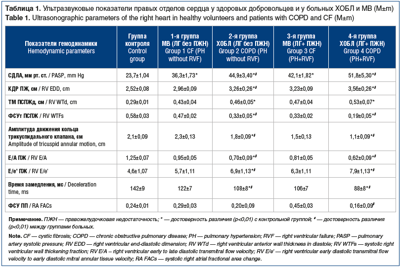
Emerging signs of myocardial diastolic dysfunction, seen in groups 1 and 2, were more significant in patients with RV failure signs (i.e., in groups 3 and 4). Moreover, these abnormalities were more significant in COPD than in CF. Thus, RV Е/А and RV Е/е' were 0.62±0.09 and 7,9±1,13, respectively, in group 4 and 0.81±0.05 and 6.3±1.11, respectively, in group 3 (р<0.01).
Therefore, in patients without significant hypoxemia, hypercapnia, and chronic intoxication, RV myocardial reserves are good enough. Meanwhile, in patients with PH, abnormal arterial blood gases, and chronic lung inflammation, RV experiences a higher load. After functional reserves have been exhausted, the chronic pulmonary heart develops [12, 13]. In addition, abnormalities in left ventricular (LV) function resulting from atrial dyssynchrony also occur. Herewith, the atrial septum functionally becomes a part of hypertrophic and dilated right ventricle, thereby reducing RV ejection fraction (EF) and resulting in LV failure [14].
These findings demonstrate that RV diastolic function (in contrast to systolic function) has lower compensation abilities which impair first [15, 16]. This impairment in RV relaxation (manifesting itself in slower pressure reduction) results in the reduction of peak early diastolic filling velocity and provokes both an increase in duration and reduction in velocity of early diastolic flow deceleration in patients with chronic lung disorders.
Changes in RV early to late diastolic transmitral flow velocity (E/A) illustrate pre- and afterload and myocardial elasticity, i.e., its stiffness and stretch, on which the value of passive ventricular filling depends. In addition, changes in time parameters during the rapid filling phase allow for calculating the contraction rate of the external oblique and internal rectus muscles. These alterations provide further ventricular shape and volume changes, wall thinning, and suction power development [17, 18]. Negative factors provoking diastolic function impairment (deceleration of relaxation, increase in RV wall stiffness) are chronic hypoxia, hypercapnia, acidosis, and anamnestic long-term chronic intoxication in COPD.
We agree with authors who believe that RV functionality is associated with the severity of arterial hypoxemia and hypercapnia, type and duration of infectious, toxic, and autoimmune effects on myocardium rather than blood pressure in pulmonary circulation [19, 20]. The trend towards an increase in some fraction and velocity myocardial indices and unchanged amplitude of tricuspid annular motion is a normal compensatory response to RV afterload growth.
There are two classic variants of chronic heart failure (CHF). Systolic CHF is diagnosed in LV EF less than 45%, and diastolic CHF is diagnosed in LV EF higher than 45%, LV diastolic dysfunction, and clinical signs of CHF. Therefore, subclinical reduction in heart contractility with the development of diastolic dysfunction and relatively preserved EF initially seen in COPD should be considered when managing these patients.
Our findings demonstrate that RV hypertrophy and failure occur in relatively low pulmonary artery pressure. In addition, hypertrophy and dilation are often concomitant in systolic PASP less than 40-50 mm Hg.
The efficacy and safety of combinations of long-term bronchodilators are also an essential issue in COPD and cardiovascular disorders. A complex analysis of several large trials has demonstrated that adding olodaterol to tiotropium is not associated with an increased risk of cardiovascular adverse events. Therefore, a combination of tiotropium and olodaterol is a suitable therapy even in the presence of cardiovascular risk factors [21]. Furthermore, retrospective analysis of tiotropium/olodaterol combination vs. monotherapy did not reveal any long-term increase in HR, systolic and diastolic BP [22]. Hence, a combination of tiotropium and olodaterol is an effective and safe treatment for COPD and comorbid cardiovascular disorders.
Conclusions
In CF, the right atrium compensates for RV dysfunction. Right atrial contractility increases in abnormal RV dysfunction and reduces only in severe decompensation of the chronic pulmonary heart. In CF, diastolic dysfunction of the right heart often precedes systolic dysfunction. More severe structural and functional abnormalities of the right ventricle are reported in COPD. Compensation abilities of the right and left ventricles exhaust, thereby providing the conditions for the progression of heart failure and the development of the chronic pulmonary heart.
Acknowledgements
The technical edition is supported by Boehringer Ingelheim.
About the authors:
Olga N. Titova — Dr. Sc. (Med.), Professor, Director of the Research Institute of Pulmonology, I.P. Pavlov First St. Petersburg State Medical University; 6–8, Lev Tolstoy str., St. Petersburg, 197022, Russian Federation; ORCID iD 0000-0003-4678-3904.
Nataliya A. Kuzubova — Dr. Sc. (Med.), Deputy Director of the Research Institute of Pulmonology, I.P. Pavlov First St. Petersburg State Medical University; 6–8, Lev Tolstoy str., St. Petersburg, 197022, Russian Federation; ORCID iD 0000-0002-1166-9717.
Albert L. Aleksandrov — Dr. Sc. (Med.), Professor, Head of Division of the Research Institute of Pulmonology, I.P. Pavlov First St. Petersburg State Medical University; 6–8, Lev Tolstoy str., St. Petersburg, 197022, Russian Federation; ORCID iD 0000-0002-9246-5256.
Vitaliy E. Perlei — Dr. Sc. (Med.), Professor, leading researcher of the Research Institute of Pulmonology, I.P. Pavlov First St. Petersburg State Medical University; 6–8, Lev Tolstoy str., St. Petersburg, 197022, Russian Federation; ORCID iD 0000-0001-6292-3888.
Aleksey Yu. Gichkin — C. Sc. (Med.), functional diagnostician of the Research Institute of Pulmonology, I.P. Pavlov First St. Petersburg State Medical University; 6–8, Lev Tolstoy str., St. Petersburg, 197022, Russian Federation; ORCID iD 0000-0003-1127-0041.
Contact information: Nataliya A. Kuzubova, e-mail: kuzubova@mail.ru.
Financial Disclosure: no authors have a financial or property interest in any material or method mentioned.
There is no conflict of interests.
Received 05.07.2021.
Revised 28.07.2021.
Accepted 20.08.2021.
2. Gorelik I.L., Kalmanova E.N., Aisanov Z.R., Chuchalin A.G. Diagnosis of early signs of cardiac remodeling in patients with COPD. Prakticheskaya meditsina. 2011;3(51):72–77 (in Russ.).
3. Kuzubova N.A. Pathophysiological mechanisms of the formation of chronic obstructive pulmonary disease (clinical and experimental study):thesis. SPb.; 2009 (in Russ.).
4. Menshikova I.G., Loskutova N.V., Sundukova E.A., Kvasnikova Yu.V. Predictors of the progression of circulatory failure in patients with chronic obstructive pulmonary disease complicated by chronic cor pulmonale. Dal’nevostochnyy meditsinskiy zhurnal. 2012;2:21–24 (in Russ.).
5. Ziegler B., Perin C., Casarotto F.C. et al. Pulmonary hypertension as estimated by Doppler echocardiography in adolescent and adult patients with cystic fibrosis and their relationship with clinical, lung function and sleep findings. Clin Respir J. 2018;12(2):754–761. DOI: 10.1111/crj.12590.
6. Tonelli A.R., Fernandez-Bussy S., Lodhi S. et al. Prevalence of pulmonary hypertension in end-stage cystic fibrosis and correlation with survival. J Heart Lung Transplant. 2010;29(8):865–872. DOI: 10.1016/j.healun.2010.04.006.
7. Podolska E., Pogorzelski A., Woś H. Echokardiograficzna ocena wystepowania serca płucnego u chorych z mukowiscydoza [Echocardiographic assessment of cor pulmonale in patients with cystic fibrosis]. Wiad Lek. 2006;59(3–4):208–213.
8. Burghuber O.C., Salzer-Muhar U., Bergmann H., Götz M. Right ventricular performance and pulmonary haemodynamics in adolescent and adult patients with cystic fibrosis. Eur J Pediatr. 1988;148(3):187–192. DOI: 10.1007/BF00441398.
9. Belle-van Meerkerk G., Cramer M.J., Kwakkel-van Erp J.M. et al. Pulmonary hypertension is a mild comorbidity in end-stage cystic fibrosis patients. J Heart Lung Transplant. 2013;32(6):609–614. DOI: 10.1016/j.healun.2013.03.006.
10. Lang R.M., Badano L.P., Mor-Avi V. et al. Recommendations for cardiac chamber quantification by echocardiography in adults: an update from the American Society of Echocardiography and the European Association of Cardiovascular Imaging. Eur Heart J Cardiovasc Imaging. 2015;16(3):233–270. DOI: 10.1093/ehjci/jev014.
11. Chen W., Thomas J., Sadatsafavi M., FitzGerald J.M. Risk of cardiovascular comorbidity in patients with chronic obstructive pulmonary disease: a systematic review and meta-analysis. Lancet Respir Med. 2015;3(8):631–639. DOI: 10.1016/S2213-2600(15)00241-6.
12. Chronic obstructive pulmonary disease: monograph / ed. prof. A.G. Chuchalin. M.: Atmosphere; 2011 (in Russ.).
13. Alter P., Watz H., Kahnert K. et al. Airway obstruction and lung hyperinflation in COPD are linked to an impaired left ventricular diastolic filling. Respir Med. 2018;137:14–22. DOI: 10.1016/j.rmed.2018.02.011.
14. Badano L.P., Ginghina C., Easaw J. et al. Right ventricle in pulmonary arterial hypertension: haemodynamics, structural changes, imaging, and proposal of a study protocol aimed to assess remodelling and treatment effects. Eur J Echocardiogr. 2010;11(1):27–37. DOI: 10.1093/ejechocard/jep152.
15. Fenster B.E., Holm K.E., Weinberger H.D. et al. Right ventricular diastolic function and exercise capacity in COPD. Respir Med. 2015;109(10):1287–1292. DOI: 10.1016/j.rmed.2015.09.003.
16. Güder G, Störk S. COPD and heart failure: differential diagnosis and comorbidity. Herz. 2019;44(6):502–508. DOI: 10.1007/s00059-019-4814-7.
17. Howard L.S., Grapsa J., Dawson D. et al. Echocardiographic assessment of pulmonary hypertension: standard operating procedure. Eur Respir Rev. 2012;21(125):239–248. DOI: 10.1183/09059180.00003912.
18. Meluzín J., Spinarová L., Bakala J. et al. Pulsed Doppler tissue imaging of the velocity of tricuspid annular systolic motion; a new, rapid, and non-invasive method of evaluating right ventricular systolic function. Eur Heart J. 2001;22(4):340–348. DOI: 10.1053/euhj.2000.2296.
19. Mocan M., Mocan Hognogi L.D., Anton F.P. et al. Biomarkers of Inflammation in Left Ventricular Diastolic Dysfunction. Dis Markers. 2019;2019:7583690. DOI: 10.1155/2019/7583690.
20. Vonk Noordegraaf A., Galiè N. The role of the right ventricle in pulmonary arterial hypertension. Eur Respir Rev. 2011;20(122):243–253. DOI: 10.1183/09059180.00006511.
21. Ferguson G.T., Buhl R., Bothner U. et al. Safety of tiotropium/olodaterol in chronic obstructive pulmonary disease: pooled analysis of three large, 52-week, randomized clinical trials. Respir Med. 2018;143:67–73. DOI: 10.1016/j.rmed.2018.08.012.
22. Andreas S., McGarvey L., Bothner U. et al. Absence of Adverse Effects of Tiotropium/Olodaterol Compared with the Monocomponents on Long-Term Heart Rate and Blood Pressure in Patients with Moderate-to-Very-Severe COPD. Int J Chron Obstruct Pulmon Dis. 2020;15:1935–1944. DOI: 10.2147/COPD.S246348.

This work is licensed under a Creative Commons «Attribution» 4.0 License.

Распечатать